Download
1 / 23
250 likes | 799 Vues
Eczema. د.سهاد الجبوري. Eczema : the ward ‘ eczema’derived from Greek ekzein, meaning to “to boil forth” or to “effervesce”. it Is a pattern of cutaneous inflammatory response .

E N D
Eczema د.سهاد الجبوري
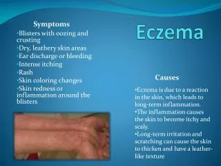
Eczema : the ward ‘eczema’derived from Greek ekzein,meaning to “to boil forth” or to “effervesce”. it Is a pattern of cutaneous inflammatory response . characterized clinically by : itching , redness , weeping in its acute form and by : dryness , lichenfication in its chronic form.
and characterized histologically by : 1- lymphocytic infiltrate 2- spongiosis ( intercellular edema ) 3- varying degrees of acanthosis ( increased thickness of epidermal layer ) 4-hyperkeratosis
Classification : 1- endogenous ( constitutional type) : a- atopic eczema ( dermatitis) b- seborrhoeic eczema (dermatitis) c- discoid eczema d- pompholyx e- stasis eczema 2- exogenous eczema (contact dermatitis): a- irritant contact dermatitis b- allergic contact dermatitis 3- unclassified : a- neurodermatitis ( lichen simplex chronicus) b- juvenial planter dermatosis
Atopic Dermatitis ( Eczema) Atopy: is a genetically determined disorder in which : 1-there is increased liability to form IgE antibodies 2-there is an increased tendency to have : asthma , hay fever & atopic dermatitis Prevalence : 10-20% of the population are affected .
Aetiology: 1- Definite aetiology are not well determined 2- AD patients usually have high level of IgE antibodies to ( house dust mites ) 3- foods clearly exacerbate symptoms in some atopic patients especially children . Eggs , nuts ,cows milk represent 75% of positive food allergies . 4- exacerbation also occurs after : immunization , viral infections ,in winter . 5- worsening factors : a- cloths irritation b- allergens of air c -excessive washing d- excessive rubbing
Clinical stages : 1- AD pass into clinicohistological evolution from : acute eczematous eruption in early life to chronic lichenified dermatitis in older patients . 2 - AD can be divided into 3 stages ( according to the onset ) : a- infantile AD 2 mo - 2 yr b- childhood AD 2 yr - 12 yr c- adolescent and adult onset AD
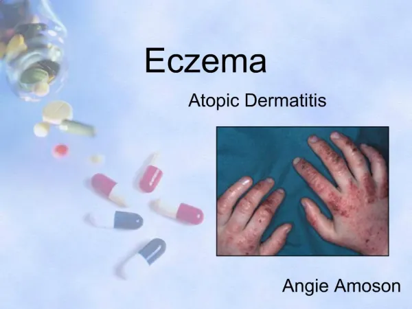
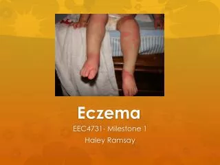
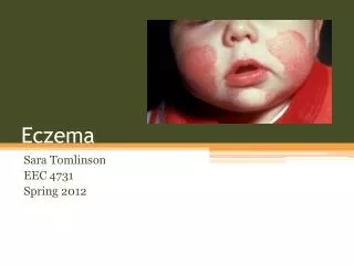
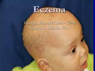
Clinical features : * in infancy presented ( mostly after 2 mo. of age )as itchy , erythema of cheeks , in these patches ,fine vesicles develop, rupture and produce moist crusted areas ( i.e. acute moist lesions ). Other sites : scalp, neck, extensor extremities , but diaper area spared . * in childhood AD , usually less exudative , drier , slightly scaly patches involving : eyelids and face , antecubitalfossae , poplitealfossae * adult AD : localized erythematous scaly papulovesicular plaques or chronic lichenified plaques, involving same sites of childhood AD .
Diagnosis : It is based on major and minor criteria and the diagnosis must be : 3 majors + 3 minors Major criteria: 1- pruritus 2- typical morphology &distribution : a- flexural lichenification in children and adults . b- facial &extensor involvement in infancy . 3- chronic or chronically relapsing dermatitis . 4- personal or family history of atopic disease ( asthma , alleric rhinitis , atopic dermatitis)
Minor criteria : 1- xerosis ( dryness ) 2- ichthyosis /hyperlinear palms/ keratosispilaris 3- increased serum IgE 4- tendency for cutaneous infection especially (staph.aureus & HSV). 5- tendency to non specific hand /foot dermatitis 6- chelitis ( inflammation of lips) 7- Dennie-Morgan infraorbital folds (> 2 folds ) 8- orbital darkening 9- facial pallor /facial erythema 10- pityriasis alba 11- perifollicular accentuation 12- white dermographism .. .. 23-nipple eczema
Immunopathology of AD: • *It is a T-helper type (Th2 dominance ) in tissues. • Th2 produce IL4 , 5 ,10 • *IL4 leads to elevated IgE & oesinophilia in tissues &peripheral blood . • * IL10 will inhibit cellular immunity • So , there is tendency towards humeral immunity. • * Langerhans cells in skin ( Ag presenting cell in skin ) are abnormal • ( directly stimulate Th cells without Ag in the way of Th2 phenotype)
Differential Diagnosis: Infantile AD should be differentiated from seborrhoeic dermatitis in infancy because of similar presentations .However , it can be differentiated by :
Treatment : 1- General measures : avoidance of:- excessive bathing ( or washing ) - extremes of cold and heat , -emotional stress , -vigorous rubbing . 2- Specific measures : A- Topical Rx : * drying agent : e.g. K+ permanganate for weeping lesions. * emollients : for hydration of dry skin e.g. Vaseline ointment and Zinc Oxide ointment . * topical steroids :( moderate to potent ) are very beneficial *topical calcineurin inhibitors :such as tacrolimus or pimecrolimus offer an alternative to topical steroids. B- Systemic Rx : 1- antihistamine : may be used for their sedative effects. 2- systemic corticosteroids to control acute and severe cases 3- Phtotherapy are often helpful for severe AD ( PUVA , NB-UVB )