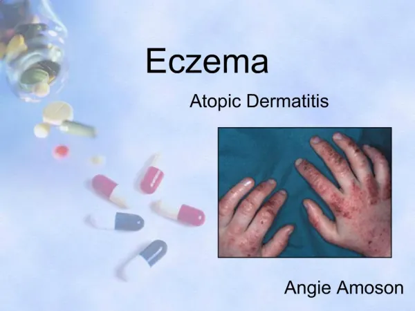
Eczema
Eczema. Definition. Eczema/dermatitis is a type of polymorphic inflammatory reaction pattern involving the epidermis and dermis which may be provoked by a number of external or internal factors and can be defined histologically by spongiosis and interstitial vesicles. Eczema’s clinical stages.

Eczema
E N D
Presentation Transcript
Definition • Eczema/dermatitis is a type of polymorphic inflammatory reaction pattern involving the epidermis and dermis which may be provoked by a number of external or internal factors and can be defined histologically by spongiosis and interstitial vesicles.
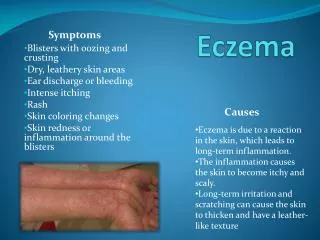
Eczema’s clinical stages • erythematous(dermatitis) • vesiculous (papulovesicular eczema) • exudative (oozing, weeping eczema) • crustification (crusted eczema) • descuamative (scaling eczema) • lichenification (lichenoid eczema)
Clinical Features • Acute eczema: it is an erythema and edema in plaques, usually with ill-defined borders. Afterwards pruritic vesicles, exudation, crusting and finally scaling. The resolution is without scars. • Chronic eczema: it is less vesicular and exudative, more scaly, pigmented and thickened, more likely to develop painful fissures and to be lichenified.
Classification • Exogenous eczemas: irritant contact dermatitis, allergic contact dermatitis; • Endogenous eczemas: atopic dermatitis; • Exo-endogenous eczemas: dyshidrotic eczema, discoid (nummular) eczema, seborrhoeic eczema (dermatitis), microbial eczema, mycotic eczema, varicose eczema, asteatotic eczema, etc.
Irritant contact eczema (dermatitis) • Irritant contact dermatitis: it is nonimmunologic local inflammatory reaction following single or repeated exposures to toxic chemicals. • a. acute irritant contact dermatitis: strong irritants produce it after a single application or a few applications. Clinical aspect: erythema, edema, vesicles or bullae, something necrosis. Reactions occur within minutes to hours after exposure to the chemical agent, are localized to the areas of maximal contact and have sharp borders. In most cases, healing appears soon after exposure. • b. chronic irritant dermatitis: it is due to repeated insults by low grade irritants. The clinical appearance ranges from dryness and cracking of the skin to erythema, vesicles, scaling, fissuring and gradual thickening. The lesions have a poor demarcation from the surrounding skin. This dermatitis may takes weeks, months or even years to develop.
Allergic contact eczema (dermatitis) • Allergic contact eczema (dermatitis): several or many exposures are usually necessary before sensitization • acute allergic contact dermatitis is characterized by erythema, edema, vesicles, exudation, crusting and scaling. Pruritus is present. • chronic allergic contact dermatitis: the skin becomes dry, scaly and thicker. Fissures and lichenification may develop later. Secondary dissemination to distant regions is frequent. • Special types: • allergic phytodermatitis • contact urticaria: nonimmunologic or allergic; • phototoxic and photoallergic contact dermatitis; • dermatitis for airborne agents et al.
Etiology of contact dermatitis • Contact irritants: alkalis, acids, phenolic compounds, amines, organic solvents, sulfur, retinoic acid, iodine, detergents, petroleum products et al. • Contact allergens: nickel, chromium, cobalt, formaldehyde resins, epoxy resins, acrylates, topical drugs, cosmetics, plants, colorants, perfumes, preservatives, et al. • Contact phyto-allergens: pentadecylcatechols (phenolic compound), which are present in poison ivy, poison oak, poison sumac, Brazilian pepper, cashew nut tree, ginkgo tree, Indian marker nut tree, lacquer tree, mango tree, rengas tree.
Pathogenesis of contact eczema • Irritant contact dermatitis: irritant substances are capable of producing a direct toxic injury and an inflammatory reaction in the skin in every person, after the contact. Immunological processes are not involved. Factors affecting the cutaneous response are: the substance’s chemical and physical properties, concentration, vehicle, duration of exposure, patient’s age, area of action, underlying dermatitis, genetic make-up, environmental factors etc. • Allergic contact dermatitis is a type IV, delayed, cell-mediated reaction. Initially, a hapten contacts the skin and forms a hapten-carrier protein complex. This complex associated in epidermal Langerhans cell, which presents this complete antigen to a T-helper cell, causing the release of various mediators. T-cell expansion in regional lymph nodes produces specific memory and T-effectors lymphocytes which circulate in the general bloodstream. The duration of process of sensitization is 5 to 21 days. Upon re-exposure to the specific antigen, a proliferation of activated T cell is produced, the mediator release and the migrations of cytotoxic T-cells, resulting in an inflammation, which appear in 48 to 72 hours after exposure.
Ag hapten Ag repeatedly ECZEMA Cytokines (IL-1, 3, 4, 6) HLA-DR CLs LT sensitized ICAM-1 VCAM-1 HLA-DR K VEC IL-1 IL-6 GM-CSF IL-1 CLS HLA-DR TCR LT-memory To skin IFN- IL-2 IFN- IL-2 IL-1 Macrophage LT memory Lymph node IFN- IL-2 IL-1 CONTACT reactivity (type IV hypersensitivity)
Atopic eczema: definition • Atopic eczema (dermatitis) is an acute, subacute or chronic relapsing skin disorder that usually begins in infancy (after first 2 months) and is characterized principally by marked pruritus, which with rubbing and scratching leads to lichenification (hyperplasia of the skin). • The serum IgE level is usually elevated. • A personal or family history of atopic eczema, allergic rhinitis and asthma is often associated.
Pathogenesis of atopic eczema • The pathogenesis of atopic eczema is complex, but involves immunological abnormalities, environmental factors and emotional influence • Immunological abnormalities in the atopic state include increased serum total IgE and specific IgE antibodies to ingested or inhaled antigens, and preferential activation of the Th2 phenotype CD4 T-cells, which form IL-4 and IL-5. • The interleukins stimulate IgE synthesis by B-cells / plasmocytes (type I hypersensitivity) • Staphylococci colonize the skin of patients with atopic eczema, and staphylococcal exotoxins with superantigen properties are also thought to play a pathogenic role in T- an B-cell abnormal activity. • It has been shown that epidermal Langerhans cells possess high affinity IgE receptors through which an eczema-like reaction (type IV hypersensitivity) could be mediated. • Both, type I and IV, hypersensitivities maintain and mutual amplify vicious course of atopic eczema.
Ag Pruritus K Asthma Urticaria Rhinitis IL-1 Ag Histamine Eo LTh0 IL-5 Mastcell IgE PL Mediators LB IL-2 IL-6 IL-4 LTh2 CLs LTs LTh LN Citokine IL-1,3,4,6 Eczema Ag ATOPIC reactivity (type I and IV hypersensitivity)
Atopic eczema: genetic aspects • The inheritance pattern is probably autosomal-dominant • 60% of adults with atopic eczema have children with atopic eczema • The prevalence in children is 80-85% when both parents had atopic eczema • HLA haplotype associations: • atopic eczema – HLA B9; • hay fever (allergic rhinitis/conjunctivitis) – HLA B12; • allergic asthma – HLA B2, B7 şi B40.
Atopic eczema: eliciting factors • Foods: eggs, milk, peanuts, soybeans, fish, and wheat • Inhalants: specific aeroallergens, especially dust mites • Microbial agents: exotoxins of Staphylococcus aureus may act as superantigens; group A streptococcus, herpes simplex virus, fungus (candidiasis and dermatophytosis) • Skin dehydration: frequent bathing and hand washing • Hormonal: pregnancy, menstruation, thyroid • Season: in temperate climates, usually improves in summer, flares in winter • Clothing: pruritus flares after taking of clothing; wool clothing or blankets directly in contact with skin • Emotional stress: primary or secondary resulting from the disease
Atopic eczema: basic clinical features(by Hanifin and Rajka) • Pruritus (if there is no scratching, there is no eruption) - itch scratch rash itch; • Typical morphology and distribution of lesions for age; • Chronic or chronically relapsing course; • Personal or family history of asthma, allergic rhinitis, or atopic eczema.
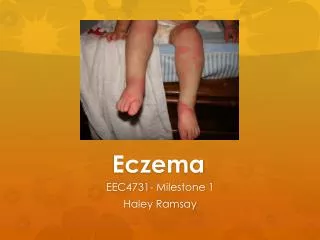
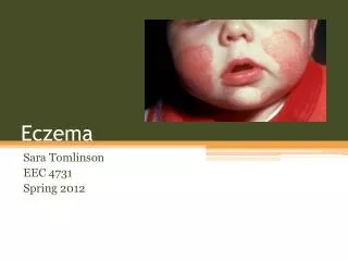
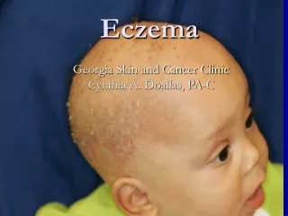
Atopic eczema at different ages • Infantile phase (2 mo – 2 yrs): characterized by intense itching, erythema, papules, vesicles, oozing and crusting; typically location on cheeks, forehead, and scalp (acute eczema). • Childhood phase (3-11 yrs): more chronic, lichenified scaly patches and plaques that may have crusting and oozing; classic areas include the wrists, ankles, backs of the thighs, buttocks, and antecubital and popliteal fossae (subacute eczema). • Adolescent/young adult phase (12-20 yrs): thick, dry, lichenified plaques that involve the face, neck, upper arms, back, and flexures (chronic or lichenified eczema). • Adult phase (>20 yrs): most commonly involves the hands, sometimes the neck and face, and rarely diffuse areas (lichenified eczema, nummular eczema, neurodermitis). Only 10% of infantile or childhood cases of atopic eczema persist into adulthood.
Atopic eczema: minor clinical features • Dry skin • Keratosis pilaris (follicular horny plugs) • Hyperlinear palms and soles • Periorbital bluish discoloration • Dennie-Morgan lines (double infraorbital fold) • Pityriasis alba (dry streptococcal skin infection) • Vascular abnormalities (skin pallor) • Cataracts • White dermatographism
Dyshidrotic eczema • Supposed association with increased sweat gland • Crops of clear, deep-seated vesicles on the palms and sides of the fingers, rarely present on soles as well • High prevalence of atopy • Sensibility to nickel, fungi, oral allergens, etc.
Nummular (discoid) eczema • Rapid onset of tiny papules and papulovesicles that form erythematous, coin-shaped plaques, ranged in size 1-10cm in diameter, resting on a background of dry skin • Most commonly occur on the extensor surfaces of the lower extremities, and often a bilaterally symmetrical, may recur at the sites of previous involvement, and are typically pruritic • It is related to dry skin, and aggravated by wool, soaps, frequent bathing, and S. aureus colonization – may be a clinical presentation of atopic eczema in adults.
Seborrheic eczema (dermatitis) • Presents in infants from 2-10 weeks until 8-12 months, and then reappears at puberty • In adults, from the beginning – dandruff; then dull or yellowish-red, sharply marginated, non-pruritic lesions covered with greasy scales on medial eyebrows, glabella, naso-labial crease, eyelid margins, post-auricular and ear canal, presternal or interscapular areas; intertriginous areas, such the inframammary crease, umbilicus, anogenital and genitocrural folds are occasionally involved • Sensitizing to Pityrosporum or Malassezia yeasts
Eczema: treatment and prevention • 1. Avoidance of as many irritants as possible; • 2. Allergen avoidance; • 3. Systemic therapy: antihistamines, glucocorticoids in severe cases; • 4. Topical treatment: • Drying agents (aluminum sulfate, calcium acetate), in acute, vesicular, weeping eruptions; • Corticosteroids: can be super, high, mid or low potent. • from superpotent corticosterids are: clobetasol dipropionate 0,05% (Dermovate), bethamethasone dipripionate 0,5% (Locacorten) et al. • high potency corticosteroids are: fluocinonide 0,05% (Cyclocort), methylprednisolone aceponate 0,1% (Advantan), mometasone furoate 0,1% (Elocom), hydrocortisone butirate 0,1% (Locoid) et al. • mid potency corticosteroids are: fluticasone propionate 0,05% (Cutivate) et al. • low potency are: hydrocortisone acetate 0,25-2,5% et al. • Solutions, lotions, gels or sprays are recommended for inflammatory, exudativ lesions and for hairy areas. • Creams and lotions are best intertriginous locations. • Ointments have good action on chronic thickened lesions.
Atopic eczema: treatment / prevention • The avoidance of irritants and allergens. • Systemic therapy: • antihistamines: sedating H1 antihistamines (promethayine, trimeprayine or hidroxyyine, alternatively) and new H1 antihistamines (cetriyine, loratadine, desloratodine, ebastine, levocetriyine, etc); • Ketotifen – a mast cell degranulation inhibitor; • Immunosupressive drugs: corticosteroids: are indicated in cases that do not respond to antihistamines, topical steroids, lubricants etc. (0,5 – 1 mg/kg/day, 7-21 days); cyclosporine A (Sandimmun or Neoral – 2-5 mg/kg/day in short-term therapy); azathioprine (Imuran, 50 mg twice daily); mycophenolate mofetil (Cellcept, 2 g/day, orally); interferon gamma; • Topical treatment • Wet-to-dry comresses for acute, inflammatory and weeping phase; • Topical corticosteroids; • Topical antibiotics • Topical immunomodulators – Tacrolimus or Pimecrolimus • Physiotherapy • PUVA • Extracorporal photopheresis • High-dose UVA therapy (340-400 nm) • Combination of UVB irradiation with UVA irradiation • Narrow-band UVB