Download
1 / 30
310 likes | 361 Vues
Dive into the physics embedded in cells with a study of biological cell structures and processes, cell theory evolution, and the significance of physics in cell biology. Learn about cell division, metabolism, and the differences between eukaryotic and prokaryotic cells.

E N D
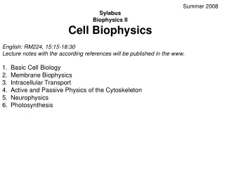
Summer 2008 • Sylabus • Biophysics II • Cell Biophysics • English: RM224, 15:15-18:30 • Lecture notes with the according references will be published in the www. • Basic Cell Biology • Membrane Biophysics • Intracellular Transport • Active and Passive Physics of the Cytoskeleton • Neurophysics • Photosynthesis
Homework 1 Is there physics in cells and in which parts of a cell is physic needed for an understanding?
Biological Cell Introduction - Cell Biology • Textbooks • Alberts B, Johnson A, Lewis J, Raff M, Roberts K, Walter P (2002). Molecular Biology of the Cell, 4th ed., Garland. ISBN 0815332181. • Lodish H, Berk A, Matsudaira P, Kaiser CA, Krieger M, Scott MP, Zipurksy SL, Darnell J (2004). Molecular Cell Biology, 5th ed., WH Freeman: New York, NY. ISBN 978-0716743668. • History • 1632 – 1723: Antonie van Leeuwenhoek teaches himself to grind lenses, builds a microscope and draws protozoa, such as Vorticella from rain water, and bacteria from his own mouth. • 1665: Robert Hooke discovers cells in cork, then in living plant tissue using an early microscope.[3] • 1839: Theodor Schwann and Matthias Jakob Schleiden elucidate the principle that plants and animals are made of cells, concluding that cells are a common unit of structure and development, and thus founding the cell theory. • The belief that life forms are able to occur spontaneously (generatio spontanea) is contradicted by Louis Pasteur (1822 – 1895) (although Francesco Redi had performed an experiment in 1668 that suggested the same conclusion). • 1855: Rudolph Virchow states that cells always emerge from cell divisions (omnis cellula ex cellula). • 1931: Ernst Ruska builds first transmission electron microscope (TEM) at the University of Berlin. By 1935, he has built an EM with twice the resolution of a light microscope, revealing previously-unresolvable organelles. • 1953: Watson and Crick made their first announcement on the double-helix structure for DNA on February 28.
Definition The cell is the structural and functional unit of all knownlivingorganisms. It is the smallest unit of an organism that is classified as living, and is sometimes called the building block of life. Some organisms, such as most bacteria, are unicellular (consist of a single cell). Other organisms, such as humans, are multicellular. (Humans have an estimated 100 trillion or 1014 cells; a typical cell size is 10 µm; a typical cell mass is 1 nanogram.) In 1837 before the final cell theory was developed, a CzechJan Evangelista Purkyně observed small "granules" while looking at the plant tissue through a microscope. The word cell comes from the Latincellula, meaning, a small room. The descriptive name for the smallest living biological structure was chosen by Robert Hooke in a book he published in 1665 when he compared the cork cells he saw through his microscope to the small rooms monks lived in. Cell Theory The cell theory, first developed in 1839 by Matthias Jakob Schleiden and Theodor Schwann, states that all organisms are composed of one or more cells. All cells come from preexisting cells. Vital functions of an organism occur within cells, and all cells contain the hereditary information necessary for regulating cell functions and for transmitting information to the next generation of cells.
All cells have several different abilities: • Reproduction by cell division: (binary fission/mitosis or meiosis). • Use of enzymes and other proteinscoded for by DNAgenes and made via messenger RNA intermediates and ribosomes. • Metabolism, including taking in raw materials, building cell components, converting energy, molecules and releasing by-products. The functioning of a cell depends upon its ability to extract and use chemical energy stored in organic molecules. This energy is released and then used in metabolic pathways. • Response to external and internal stimuli such as changes in temperature, pH or levels of nutrients. • Cell contents are contained within a cell surface membrane that is made from a lipid bilayer with proteins embedded in it. There are two types of cells: eukaryotic and prokaryotic. Prokaryotic cells are usually independent, while eukaryotic cells are often found in multicellular organisms.
Prokaryotic cells • Prokaryotes differ from eukaryotes since they lack of a nuclear membrane and a cell nucleus. Prokaryotes also lack most of the intracellular organelles and structures that are seen in eukaryotic cells. There are two kinds of prokaryotes, bacteria and archaea, but these are similar in the overall structures of their cells. Most functions of organelles, such as mitochondria, chloroplasts, and the Golgi apparatus, are taken over by the prokaryotic cell's plasma membrane. Prokaryotic cells have three architectural regions: appendages called flagella and pili — proteins attached to the cell surface; a cell envelope - consisting of a capsule, a cell wall, and a plasma membrane; and a cytoplasmic region that contains the cell genome (DNA) and ribosomes and various sorts of inclusions. • Other differences include: • The plasma membrane (a phospholipid bilayer) separates the interior of the cell from its environment and serves as a filter and communications beacon. • Most prokaryotes have a cell wall (some exceptions are Mycoplasma (bacteria) and Thermoplasma (archaea)). This wall consists of peptidoglycan in bacteria, and acts as an additional barrier against exterior forces. It also prevents the cell from "exploding" (cytolysis) from osmotic pressure against a hypotonic environment. A cell wall is also present in some eukaryotes like plants (cellulose) and fungi, but has a different chemical composition. • A prokaryotic chromosome is usually a circular molecule (an exception is that of the bacterium Borrelia burgdorferi, which causes Lyme disease). Even without a real nucleus, the DNA is condensed in a nucleoid. Prokaryotes can carry extrachromosomal DNA elements called plasmids, which are usually circular. Plasmids can carry additional functions, such as antibiotic resistance.
Eukaryotic cells • Eukaryotic cells are about 10 times the size of a typical prokaryote and can be as much as 1000 times greater in volume. The major difference between prokaryotes and eukaryotes is that eukaryotic cells contain membrane-bound compartments in which specific metabolic activities take place. Most important among these is the presence of a cell nucleus, a membrane-delineated compartment that houses the eukaryotic cell's DNA. It is this nucleus that gives the eukaryote its name, which means "true nucleus." • Other differences include: • The plasma membrane resembles that of prokaryotes in function, with minor differences in the setup. Cell walls may or may not be present. • The eukaryotic DNA is organized in one or more linear molecules, called chromosomes, which are associated with histone proteins. All chromosomal DNA is stored in the cell nucleus, separated from the cytoplasm by a membrane. Some eukaryotic organelles also contain some DNA. • Eukaryotes can move using cilia or flagella. The flagella are more complex than those of prokaryotes.
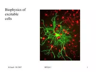
Cell Specialisation Cells can become specialised to perform a particular function within an organism, usually as part of a larger tissue consisting of many of the same cells working in tandem, for example; Nerve cells to operate as part of the nervous system to send messages back and forth via the brain at the centre of the nerve system. Skin cells for waterproof protection and protection against pathogens in the open air environment. Cells combine their efforts in these tissue types to perform a common cause. The task of the specialised cell will determine in what way it is going to be specialised, because different cells are suited to different purposes, as illustrated in the above list and below example; Muscle cells are long and smooth in structure and their elastic nature allows these cells to perform flexible movements, just as they do in our own body's. Some white blood cells contain powerful digestive enzymes to eliminate pathogens by breaking them down to the molecular level. Cells at the back of the eye are sensitive to light stimuli, and thus can interpret differences in light intensity which can in turn be interpreted by our nervous system and brain. Many of these cells contain organelles, though after some cells are specialised, they do not possess particular characteristics as they do not require them to be there. i.e. efficiency is the key, no resources are wasted and the resources available are put to their idyllic optimum.
Diagram of a typical eukaryotic cell, showing subcellular components. Organelles: (1) nucleolus(2) nucleus(3) ribosome(4) vesicle(5) rough endoplasmic reticulum (ER)(6) Golgi apparatus(7) Cytoskeleton(8) smooth ER(9) mitochondria(10) vacuole(11) cytoplasm(12) lysosome(13) centrioles within centrosome
Cell membrane: A cell's defining boundary • The cytoplasm of a cell is surrounded by a plasma membrane. The plasma membrane in plants and prokaryotes is usually covered by a cell wall. This membrane serves to separate and protect a cell from its surrounding environment and is made mostly from a double layer of lipids (hydrophobic fat-like molecules) and hydrophilicphosphorus molecules. Hence, the layer is called a phospholipid bilayer. It may also be called a fluid mosaic membrane. Embedded within this membrane is a variety of protein molecules that act as channels and pumps that move different molecules into and out of the cell. The membrane is said to be 'semi-permeable', in that it can either let a substance (molecule or ion) pass through freely, pass through to a limited extent or not pass through at all. Cell surface membranes also contain receptor proteins that allow cells to detect external signalling molecules such as hormones.
Cytoskeleton: A cell's scaffold • The cytoskeleton acts to organize and maintain the cell's shape; anchors organelles in place; helps during endocytosis, the uptake of external materials by a cell, and cytokinesis, the separation of daughter cells after cell division; and moves parts of the cell in processes of growth and mobility. The eukaryotic cytoskeleton is composed of microfilaments, intermediate filaments and microtubules. There is a great number of proteins associated with them, each controlling a cell's structure by directing, bundling, and aligning filaments. The prokaryotic cytoskeleton is less well-studied but is involved in the maintenance of cell shape, polarity and cytokinesis.
5 m Actin filaments / Microfilaments Around 7 nm in diameter, this filament is composed of two intertwined actin chains. Microfilaments are most concentrated just beneath the cell membrane, and are responsible for resisting tension and maintaining cellular shape, forming cytoplasmatic protuberances (like pseudopodia and microvilli- although these by different mechanisms), and participation in some cell-to-cell or cell-to-matrix junctions. In association with these latter roles, microfilaments are essential to transduction. They are also important for cytokinesis (specifically, formation of the cleavage furrow) and, along with myosin, muscular contraction. Actin/Myosin interactions also help produce cytoplasmic streaming in most cells. Intermediate filaments These filaments, 8 to 12 nanometers in diameter, are more stable (strongly bound) than actin filaments, and heterogeneous constituents of the cytoskeleton. Like actin filaments, they function in the maintenance of cell-shape by bearing tension (microtubules, by contrast, resist compression. It may be useful to think of micro- and intermediate filaments as cables, and of microtubules as cellular support beams). Intermediate filaments organize the internal tridimensional structure of the cell, anchoring organelles and serving as structural components of the nuclear lamina and sarcomeres. They also participate in some cell-cell and cell-matrix junctions. Microtubules Microtubules are hollow cylinders about 25 nm in diameter (lumen = approximately 15nm in diameter), most commonly comprised of 13 protofilaments which, in turn, are polymers of alpha and beta tubulin. They have a very dynamic behaviour, binding GTP for polymerization. They are commonly organized by the centrosome. They play key roles in: intracellular transport (associated with dyneins and kinesins, they transport organelles like mitochondria or vesicles), the axoneme of cilia and flagella, the mitotic spindle, and synthesis of the cell wall in plants.
Genetic material Two different kinds of genetic material exist: deoxyribonucleic acid (DNA) and ribonucleic acid (RNA). Most organisms use DNA for their long-term information storage, but some viruses (e.g., retroviruses) have RNA as their genetic material. The biological information contained in an organism is encoded in its DNA or RNA sequence. RNA is also used for information transport (e.g., mRNA) and enzymatic functions (e.g., ribosomal RNA) in organisms that use DNA for the genetic code itself. Prokaryotic genetic material is organized in a simple circular DNA molecule (the bacterial chromosome) in the nucleoid region of the cytoplasm. Eukaryotic genetic material is divided into different, linear molecules called chromosomes inside a discrete nucleus, usually with additional genetic material in some organelles like mitochondria and chloroplasts (see endosymbiotic theory). A human cell has genetic material in the nucleus (the nuclear genome) and in the mitochondria (the mitochondrial genome). In humans the nuclear genome is divided into 46 linear DNA molecules called chromosomes. The mitochondrial genome is a circular DNA molecule separate from the nuclear DNA. Although the mitochondrial genome is very small, it codes for some important proteins. Foreign genetic material (most commonly DNA) can also be artificially introduced into the cell by a process called transfection. This can be transient, if the DNA is not inserted into the cell's genome, or stable, if it is.
Organelles • The human body contains many different organs, such as the heart, lung, and kidney, with each organ performing a different function. Cells also have a set of "little organs," called organelles, that are adapted and/or specialized for carrying out one or more vital functions. Membrane-bound organelles are found only in eukaryotes
Cell nucleus (a cell's information center) • The cell nucleus is the most conspicuous organelle found in a eukaryotic cell. It houses the cell's chromosomes, and is the place where almost all DNA replication and RNAsynthesis occur. The nucleus is spherical in shape and separated from the cytoplasm by a double membrane called the nuclear envelope. The nuclear envelope isolates and protects a cell's DNA from various molecules that could accidentally damage its structure or interfere with its processing. During processing, DNA is transcribed, or copied into a special RNA, called mRNA. This mRNA is then transported out of the nucleus, where it is translated into a specific protein molecule. In prokaryotes, DNA processing takes place in the cytoplasm.
Mitochondria and Chloroplasts (the power generators) • Mitochondria are self-replicating organelles that occur in various numbers, shapes, and sizes in the cytoplasm of all eukaryotic cells. As mitochondria contain their own genome that is separate and distinct from the nuclear genome of a cell, they play a critical role in generating energy in the eukaryotic cell, they give the cell energy by the process of respiration, adding oxygen to food (typicially pertaining to glucose and ATP) to release energy. Organelles that are modified chloroplasts are broadly called plastids, and are often involved in storage. Since they contain their own genome, they are thought to have once been separate organisms, which later formed a symbiotic relationship with the cells. Chloroplasts are the counter-part of the mitochondria. Instead of giving off CO2 and H2O Plants give off glucose, oxygen, 6 molecules of water (compared to 12 in respiration) this process is called photosynthesis.
Biological Energy - ADP & ATP - Cell Biology ATP stands for Adenosine Tri-Phosphate, and is the energy used by an organism in its daily operations. It consists of an adenosine molecule and three inorganic phosphates. After a simple reaction breaking down ATP to ADP, the energy released from the breaking of a molecular bond is the energy we use to keep ourselves alive. ATP to ADP - Energy Release This is done by a simple process, in which one of the phosphate molecules is broken off, therefore reducing the ATP from 3 phosphates to 2, forming ADP (Adenosine Diphosphate after removing one of the phosphates {Pi}). This is commonly wrote as ADP + Pi. When the bond connecting the phosphate is broken, energy is released. While ATP is constantly being used up by the body in its biological processes, the energy supply can be bolstered by new sources of glucose being made available via eating food which is then broken down by the digestive system to smaller particles that can be utilised by the body. On top of this, ADP is built back up into ATP so that it can be used again in its more energetic state. Although this conversion requires energy, the process produces a net gain in energy, meaning that more energy is available by re-using ADP+Pi back into ATP. Glucose and ATP Many ATP are needed every second by a cell, so ATP is created inside them due to the demand, and the fact that organisms like ourselves are made up of millions of cells. Glucose, a sugar that is delivered via the bloodstream, is the product of the food you eat, and this is the molecule that is used to create ATP. Sweet foods provide a rich source of readily available glucose while other foods provide the materials needed to create glucose. This glucose is broken down in a series of enzyme controlled steps that allow the release of energy to be used by the organism. This process is called respiration. Respiration and the Creation of ATP ATP is created via respiration in both animals and plants. The difference with plants is the fact they attain their food from elsewhere (see photosynthesis). In essence, materials are harnessed to create ATP for biological processes. The energy can be created via cell respiration. The process of respiration occurs in 3 steps (when oxygen is present): Glycolysis The Kreb's Cycle The Cytochrome System
Intracellular Transport There are two methods : Active Transport - Active transport with the help of a molecular motor. Passive Transport (Diffusion) - The movement of molecules from areas of high concentration to areas of low concentration.Besides normal diffusion subdiffusion may occurr.
Endoplasmic reticulum and Golgi apparatus (macromolecule managers) • The endoplasmic reticulum (ER) is the transport network for molecules targeted for certain modifications and specific destinations, as compared to molecules that will float freely in the cytoplasm. The ER has two forms: the rough ER, which has ribosomes on its surface, and the smooth ER, which lacks them. Also the Golgi apparatus's ends "pinch" off and become new vacuoles in the animal cell. Note: This function is also in parts fullfilled by the cytoskeleton!
Ribosomes (the protein production centers in the cell) • Ribosomes (from ribonucleic acid and "Greek: soma (meaning body)") are complexes of RNA and protein that are found in all cells. Prokaryotic ribosomes from archaea and bacteria are smaller than most of the ribosomes from eukaryotes such as plants and animals. However, the ribosomes in the mitochondrion of eukaryotic cells resemble those in bacteria, reflecting the evolutionary origin of this organelle. • The function of ribosomes is the assembly of proteins, in a process called translation. Ribosomes do this by catalysing the assembly of individual amino acids into polypeptide chains; this involves binding a messenger RNA and then using this as a template to join together the correct sequence of amino acids. This reaction uses adapters called transfer RNA molecules, which read the sequence of the messenger RNA and are attached to the amino acids.
Lysosome Lysosomes are organelles that contain digestive enzymes (acid hydrolases). They digest excess or worn-out organelles, food particles, and engulfed viruses or bacteria. The membrane surrounding a lysosome allows the digestive enzymes to work at the 4.5 pH they require. Lysosomes fuse with vacuoles and dispense their enzymes into the vacuoles, digesting their contents. They are created by the addition of hydrolytic enzymes to early endosomes from the Golgi apparatus. The name lysosome derives from the Greek words lysis, which means dissolution or destruction, and soma, which means body. They are frequently nicknamed "suicide-bags" or "suicide-sacs" by cell biologists due to their role in autolysis. Lysosomes were discovered by the Belgian cytologist Christian de Duve in 1949. Peroxisome Peroxisomes are ubiquitousorganelles in eukaryotes that participate in the metabolism of fatty acids and other metabolites. Peroxisomes have enzymes that rid the cell of toxic peroxides. They have a single lipid bilayer membrane that separates their contents from the cytosol (the internal fluid of the cell) and contain membrane proteins critical for various functions, such as importing proteins into the organelles and aiding in proliferation. Like lysosomes, peroxisomes are part of the secretory pathway of a cell, but they are much more dynamic and can replicate by enlarging and then dividing. Peroxisomes were identified as cellular organelles by the Belgian cytologist Christian de Duve in 1967 after they had been first described in a Swedish PhD thesis a decade earlier.
Centrosome (the cytoskeleton organiser) • The centrosome produces the microtubules of a cell - a key component of the cytoskeleton. It directs the transport through the ER and the Golgi apparatus. Centrosomes are composed of two centrioles, which separate during cell division and help in the formation of the mitotic spindle. A single centrosome is present in the animal cells. They are also found in some fungi and algae cells. • Vacuoles • Vacuoles store food and waste. Some vacuoles store extra water. They are often described as liquid filled space and are surrounded by a membrane. Some cells, most notably Amoeba, have contractile vacuoles, which are able to pump water out of the cell if there is too much water.
Cell functions • Cell metabolism • Between successive cell divisions, cells grow through the functioning of cellular metabolism. • Cell metabolism is the process by which individual cells process nutrient molecules. Metabolism has two distinct divisions: catabolism, in which the cell breaks down complex molecules to produce energy and reducing power, and anabolism, in which the cell uses energy and reducing power to construct complex molecules and perform other biological functions. Complex sugars consumed by the organism can be broken down into a less chemically-complex sugar molecule called glucose. Once inside the cell, glucose is broken down to make adenosine triphosphate (ATP), a form of energy, via two different pathways. • The first pathway, glycolysis, requires no oxygen and is referred to as anaerobic metabolism. Each reaction is designed to produce some hydrogen ions that can then be used to make energy packets (ATP). In prokaryotes, glycolysis is the only method used for converting energy. • The second pathway, called the Krebs cycle, or citric acid cycle, occurs inside the mitochondria and is capable of generating enough ATP to run all the cell functions.
Cell Division Cell division involves a single cell (called a mother cell) dividing into two daughter cells. This leads to growth in multicellular organisms (the growth of tissue) and to procreation (vegetative reproduction) in unicellular organisms. Prokaryotic cells divide by binary fission. Eukaryotic cells usually undergo a process of nuclear division, called mitosis, followed by division of the cell, called cytokinesis. A diploid cell may also undergo meiosis to produce haploid cells, usually four. Haploid cells serve as gametes in multicellular organisms, fusing to form new diploid cells. DNA replication, or the process of duplicating a cell's genome, is required every time a cell divides. Replication, like all cellular activities, requires specialized proteins for carrying out the job. Schematic of the cell cycle. outer ring: I=Interphase, M=Mitosis; inner ring: M=Mitosis, G1=Gap 1, G2=Gap 2, S=Synthesis; not in ring: G0=Gap 0/Resting. The duration of mitosis in relation to the other phases has been exaggerated in this diagram
Prophase: The two round objects above the nucleus are the centrosomes. Note the condensed chromatin. Prometaphase: The nuclear membrane has degraded, and microtubules have invaded the nuclear space. These microtubules can attach to kinetochores or they can interact with opposing microtubules. Metaphase: The chromosomes have aligned at the metaphase plate. Telophase: The decondensing chromosomes are surrounded by nuclear membranes. Note cytokinesis has already begun, the pinching is known as the cleavage furrow. Early anaphase: Kinetochore microtubules shorten
Protein synthesis • Cells are capable of synthesizing new proteins, which are essential for the modulation and maintenance of cellular activities. This process involves the formation of new protein molecules from amino acid building blocks based on information encoded in DNA/RNA. Protein synthesis generally consists of two major steps: transcription and translation. • Transcription is the process where genetic information in DNA is used to produce a complementary RNA strand. This RNA strand is then processed to give messenger RNA (mRNA), which is free to migrate through the cell. mRNA molecules bind to protein-RNA complexes called ribosomes located in the cytosol, where they are translated into polypeptide sequences. The ribosome mediates the formation of a polypeptide sequence based on the mRNA sequence. The mRNA sequence directly relates to the polypeptide sequence by binding to transfer RNA (tRNA) adapter molecules in binding pockets within the ribosome. The new polypeptide then folds into a functional three-dimensional protein molecule.
Cell movement or motility Cell has the ability to move spontaneously during the process of wound healing, immune response, neuronal development and cancer metastasis. For wound healing to occur, white blood cells and cells that ingest bacteria move to the wound site to kill the microorganisms that cause infection. A the same time fibroblasts (connective tissue cells) move there to remodel damaged structures. In the case of tumor development, cells from a primary tumor move away and spread to other parts of the body. Cell motility involves many receptors, crosslinking, bundling, binding, adhesion, motor and other proteins. The process is divided into three steps - protrusion of the leading edge of the cell, adhesion of the leading edge and deadhesion at the cell body and rear, and cytoskeletal contraction to pull the cell forward. Each of these steps is driven by physical forces generated by unique segments of the cytoskeleton.
Cell Motility Moving keratozyte Moving fibroblast Moving growth cone Lamellipodium is an independent functional cellular module! Moving fragment 10 µm
Homework 2 • Find as many material constants! • Estimate the maximal size of a cell due to diffusion!