Download
1 / 42
650 likes | 3.94k Vues
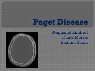
Paget’s Disease (Osteitis Deformans). Excess of bone destruction & unorganized bone formation and repair. The 2 nd most common bone disorder in the U.S. The etiology is unknown

E N D
Paget’s Disease (Osteitis Deformans) • Excess of bone destruction & unorganized bone formation and repair. The 2nd most common bone disorder in the U.S. • The etiology is unknown • Usually affects the axial skeleton, vertebrae and skull, although the pelvis, tibia, femur are the other common sites of disease. • Most persons are asymptomatic & diagnosis is incidental.
Paget’s Disease (Osteitis Deformans) • Vascularity is increased in affected portions of the skeleton. Lesions may occur in one or more bones, does not spread from bone to bone. • Deformities & bony enlargement often occur. Bowing of the limbs & spinal curvature in persons with advanced disease. • Bone pain- is the most common symptom. Is is usually worse with ambulation or activity but may also occur at rest. Involved bones may feel spongy & warm because of increased vascularity. • Skull pain is usually accompanied with headache, warmth, tenderness & enlargement of the head.
Paget’s Disease (Osteitis Deformans • Pathologic fractures- because of the increased vascularity of the involved bone-bleeding is a potential danger. • Alkaline phosphatase levels- markedly elevated as the result of osteoblast activity. • Serum calcium are normal except with generalized disease or immobilization. • Gout and hyperurecemia may develop as a result of increased bone activity, which causes an increase in nucleic acid catabolism.
Paget’s Disease (Osteitis Deformans • Radiograph reveals radiolucent areas in the bone, typical of increased bone resorption. Deformities & fractures may also be present. • Goals of the treatment- to relieve pain & prevent fracture & deformities. • Pharmacologic agents are used to suppress osteoclastic activity. Bisphosphonates & calcitonin are effective agents to decrease bone pain & bone warmth & also relieve neural decompression, joint pain & lytic lesions. • Use of analgesics & NSAIDs. Assistive devices, including cane, walker.
Paget’s Disease (Osteitis Deformans • Deformities may be corrected by surgical intervention (osteotomy). ORIF may be necessary for fractures. • The patient may benefit from a PT referral. Local application of ice or heat may help alleviate pain. • A regular exercise should be maintained; walking is best. Avoid extended periods of immobility to avoid hypercalcemia. • A nutritionally adequate diet is recommended. Assistance in learning to use canes or other ambulatory aids. • The Arthritis Foundation & Paget Foundation are useful resources for patients & their families.
Osteomyelitis • Infection of the bone, most often of the cortex or medullary portion. Is is commonly caused by bacteria, fungi, parasites & viruses. • Classified by mode of entry- Contiguous orexogenous is caused by a pathogen from outside the body or the by the spread of infection from adjacent soft tissues. The organism is Staph aureus. Example- pathogens from open fracture. The onset is insidious: initially cellulites progressing ti underlying bone.
Osteomyelitis • Hematogenous- caused by bloodborne pathogens originating from infectious sites within the body.Ex: sinus, ear, dental, respiratory & GU infections. The infection spreads from the bone to the soft tissues & can eventually break through the skin, becoming a draining fistula. Again, Staph aureus is the most common causative organism. • Acute Osteomyelitis left untreated or unresolved after 10 days is considered chronic. Necrotic bone is the distinguishing feature of chronic osteomyelitis.
Osteomyelitis • The pathophysiology is similar to that infectious processes in any other body tissue. • Bone inflammation is marked by edema, increased vascularity & leukocyte activity. • The patient report fever, malaise, anorexia, & headache. The affected body may be erythematous, tender, & edematous. There may be fistula draining purulent material. • Blood test- increase WBCs, ESR, & C-protein levels.
Osteomyelitis • Treatment is difficult & costly. The goal are complete removal of dead bone & affected soft tissue, control of infection & elimination of dead space (after removal of necrotic bone). • The nursing management-use of aseptic technique during dressing changes. Observed for S/S of systemic infection, & administered antibiotic on time. • ROM exercises are encouraged to prevent contractures & flexion deformities & participation in ADL to the fullest extent is encouraged.
Tumor of the MSS • MSS constitute 3% of all malignant tumors. • Malignant tends to cause more bone destruction, invasion of the surrounding tissues & metastasis. • Benign tumors- tend to be less destructive to normal bone. • The cause of bone tumors is unknown. • The tumor is defined as a new growth or hyperplasia of cells. This growth is in response to inflammation or trauma.
Tumor of the MSS • Osteosarcoma- A malignant tumor originating from osteoblast (bone-forming cells). Occurs twice as frequently in males as in females. • Usually located at the end of the long bones (metaphysis). Most frequently seen at the distal end of the femur or the proximal end of the tibia. • Lungs, a common site of metastasis. • Pain and swelling at the site & limitation of movement. • Bone biopsy is used to confirm the diagnosis. • X-ray films, CT scans, MRI & bone scans show tumor location & size. • Historically, the treatment of choice is amputation.
Tumor of the MSS • Ewing’s sarcoma- A malignant tumor of the bone originating from myeloblasts with early metastases to lung, lymph nodes, & other bones. • Usually located on the shaft of the long bones. Femur, tibia, & humerus are common sites. • Poor prognosis. Common in person> 40 years old. Affect males more than females. • Pain increased with weight bearing. May complain of weight loss, malaise, or anorexia. • Causes pathologic fractures. • Treatment: Palliative, radiation, chemotherapy.
Tumor of the MSS • Chondrosarcoma- Usually affects persons 50-70 years old. Accounts for 20% of all bone tumors • Affect males than females. • Slow growing, insidious onset. Most common in humerus, femur, pelvis. • Localized pain, swelling. May have palpable mass. Severe, persistent pain. May infiltrate joint space & soft tissue & metastasize to the lungs. • Treatment: Surgical incision, amputation.
Tumor of the MSS • Fibrosarcoma- Usually affects persons 30- 50 years old. Affects females than males. • Occurs in bony fibrous tissue of femur & tibia. • Accounts for 4% of primary malignant bone tumors. • May result from radiation therapy, paget’s disease or chronic osteomyelitis. • Night pain, swelling, possible palpable mass. May cause pathologic fractures. • May metastasize to the lungs.
Fractures • A break in the continuity of bone caused by trauma, twisting or as a result of bone decalcification. Results when the bone is unable to absorb the stress. Result of an accident or injury, stress fracture occur as a result of normal activity or after minimal injury. • Causes of Fracture: Fatigue- muscles are less supportive to bone, therefore, can’t absorb the force being exerted. Bone neoplasms- cellular proliferations of malignant cells replace normal tissue causing weakened bone. Metabolic disorders-poor mineral absorption & hormonal changes decreases bone calcification which results in a weakened bone. Bedrest or disuse- atropic muscles & osteoporosis causes decreased stress resistance.
Fractures (types) • Greenstick- A crack; bending of a bone with incomplete fracture. Only affects one side of the periosteum. Common in skull fractures or in young children when bones are pliable. • Comminuted: Bone completely broken in a transverse, spiral or oblique direction ( indicates the direction of the fracture in relation to the long axis of the fracture bone). Bone broken into several fragments. • Open or compound: Bone is exposed to the air through break in the skin. Can be associated with soft tissue injury. Infection is common complication due to exposure to bacterial invasion.
Fractures (types) • Closed or simple: Skin remains intact. Chances are greatly decreased for infection. • Compression: Frequently seen with vertebral fractures. Bone has been compressed by other bones. • Complete: Bone is broken with disruption of both sides of the periosteum. • Impacted: one part of fractured bone is driven into another. • Depressed: Usually seen in skull or facial fractures. Bone or fragments of bone are driven inward. • Pathological: break caused by disease process. • Intracapsular:Bone broken inside the joint. • Extracapsular: Fracture outside the joint.
Fractures (stages of bone healing) • Occurs over several weeks • New bone tissue occurs in region of break • Repair is initiated by migration of blood vessels and connective tissue from periosteum in break area. • Dense fibrous tissue fills from periosteum in break area. Osteoblast near the broken area. Chondroblast further away from broken area. • Cells deposit cartilage between broken surfaces • Cartilage is slowly replaced by mineralized bone tissue, which completes repair.
Fractures (signs & symptoms) • Pain or tenderness over involved area. • Loss of function of the extremity • Crepitation: sound of grating bone fragments • Ecchymosis or erythema • Edema • Muscle spasm • Deformity: Overriding; Angulation- limb is in unnatural position.
Fractures (Emergency care) • Immobilize affected extremity to prevent further damage to soft tissue or nerve. • If compound fracture is evident, don’t attempt to reduce it. Apply splint. Cover open wound with sterile dressing. • Use splint: External support is applied around a fracture area to immobilize the broken ends. Material used: wood, plastic (air splints), magazines. • Function of the splinting: Prevent additional trauma, reduce pain, decrease muscle spasm, limit movement, prevent complications, such as fat emboli if long bone fracture. • Provide specific care for fracture treatment: traction, cast. Surgical intervention.
Traction • Force applied in two directions to reduce and/ or immobilize a fracture, to provide proper bone alignment and regain normal length, or to reduce muscle spasm. • Closed reduction: Manual manipulation. Usually done under anesthesia to reduce pain & relax muscles, thereby preventing complications. Cast is usually applied following closed reduction. • Open reduction: Surgical intervention. Usually treated with internal fixation devices (screws, plates, wires). Cast application.
Traction • Skeletal Traction: Mechanical applied to bone, using pins (Steinmann), wires (Kirscher), or tongs (Crutchfield). Most often used in fractures of femur, tibia, humerus. • Skin traction: applied by use of elastic bandages, moleskin strips, or adhesive. Used most often in alignment or lengthening (for congenital hip displacement) or to relieve muscle spasms in preop hip clients. Most common types are: Russell, Buck’s, Cervical (used for whiplashes & cervical spasm), -pull is exerted on one plane & used for temporary immobilization; Pelvic traction (used for low back pain).
Traction(Principles) • The line of pull must be maintained. Center the patient in the bed & place in good alignment. • The pull of traction must be continuous. Remove or add weights only with MD order. • The ropes & weights must be free of friction. Be certain the weights hangs free at all times & that the ropes are over the center of the pulley. • There must be sufficient countertraction maintained at all times. Keep the patient from sliding to the foot of the bed.
Cast Care • After application of cast, allow 24 to 48 hours for drying. For synthetic cast, allow 30 minutes • Cast will change from dull to shiny substance when dry. • Don’t handle cast during dying process, because indentation from fingermarks can cause skin breakdown under cast. • Keep extremity elevated to prevent edema. • Provide for smooth edges surrounding cast. Smooth edges prevent crumbling and breaking down of edges. Stockinet can be pulled over edge & fastened down with adhesive tape to outside of cast. • Observe casted extremity for signs of circulatory impairment. Cast may have to be cut if edematous condition continues. • Always observe for sign & symptoms of complications: pain swelling, discolaration, tingling or numbness, diminished or absent pulse, paralysis, cool to touch.
Cast Care • If there is an open, draining area on the affected extremity, a window (cut out portion of cast) can be utilized for observation and/or irrigation of wound. • Keep cast dry. Breaks down when water comes in contact with plaster. Use plastic bags or Chux during bath or when using bedpan, to protect cast material. • Utilize isometric exercises to prevent muscle atrophy & to strengthen the muscle. Isometrics prevent joint from being immobilized. • Position client with pillows to prevent strain on unaffected areas. • Turn every 2 hours to prevent complications. Encourage to lie on abdomen 4 hours a day.
Cast (complications) • Respiratory complications: have client cough & DB q 2 hours. Turn q 2 hours if not contraindicated. • Thrombus & embolic formation. Apply SCD. Start anticoagulation therapy if needed. Observe for S/S of pulmonary and/or fat emboli. • Contractures: Start ROM exercises to affected joints. Provide foot board. • Skin breakdown- massage with lotion once a day to prevent drying. Alternate pressure mattress, sheepskin. Use stryker boots or heel protectors. • Prevent urinary retention and calculi. Encourage fluids. Monitor intake & output.
Cast (complication) • Prevent constipation: Encourage fluids, Provide high-fiber diet. Administer laxative or enema as ordered. • Provide psychological support: Allow to ventilate feelings of dependence. Encourage independence when possible. Encourage visitors for short time periods. Provide diversionary activities.
Fractures (Complications) • Compartment Syndrome: An increase in the pressure within the a fascial muscle compartment • Tissue damage can occur within 30 minutes & elevated pressure for more than 4 hours can result in irreversible damage & limb loss. • Signs & symptoms: 6 P’s. Pain-severe, unrelenting, unrelieved by analgesia & increased by elevation of the extremity. Pallor- coolness, slow capillary refill. Pulselessness-diminished or absent pulses. Increase pressure and paresthesia & paralysis. • Goals of treatment: decreasing tissue pressure, restoring blood flow & preserving function of the limb. • Diagnosis: Intracompartment pressure > 30 mm hg.
Fractures (Complications) • Treatment: Fasciotomy-open the affected compartment, decrease the pressure & restore normal perfusion. The wound is covered with wet saline dressing. • Fat embolism syndrome (FES)-fat globules & tissue thromboplastin are released from the bone marrow. The fat molecules enter the venous circulation, travel to the lungs & embolize the small capillaries & arterioles. • S/S: hypoxemia, tachypnea, fever, chest pain altered mental status.The presence of unexplained fever, accompanied by a change in mental status & petechiae, shld. Alert the caregiver to the possibility of FES. The MD shld. Be notified immediately.
Fractures (Complications) • Infection: leading cause of delayed union & nonunion, occurs primarily in open or compound fractures. The most symptoms occur within 4 weeks of the injury. • S/S: Pain, erythema & edema.
HIP fractures • High incidence in elderly group-most common cause of traumatic death after age 75. • Fractures caused by brittle bones (osteoporosis) 7 frequent falls in the elderly. • Elderly with hip fractures frequently have associated medical conditions (CAD, renal disorders). • Assessment: Intracapsular-bone broken inside the joint-treated by internal fixation-replacement of femoral head with Austin Moore prosthesis. • Placed in skin traction first for immobilization & relief of muscle spasm.
HIP fractures • Extracapsular: trochanteric fracture outside the joint. • Can be treated by balanced suspension traction. Full weight-bearing usually in 6 to 8 weeks, when healing takes place. • Surgery: usually internal fixation with wire. • Intertrochanteric fracture: extends from medial region of the junction of the neck & lesser trochanter toward the summit of the greater trochanter. Treated initially with balanced suspension traction. Internal fixation used with nailplate, screws & wire. Not allowed to flex hip to the side, on the side of the bed, or in a low chair. When hip is flexed, displacement can occur.
Total hip replacement • Replacement of both the acetabulum & the head of the femur with metal or plastic implants. • Used in degenerative diseases or when fracture of the head of femur has occurred with nonunion. • To prevent flexion, keep operative leg in abduction by use of pillow or abductor splints. • Keep hemovac in place until drainage has subsided (24 to 96 hours). • Prevent edema: readjust SCD at least every 4 to 8 hours. • Prevent infections- monitor prophylactic antibiotic.
HIP fractures • Continuous passive motion (CPM) first day postop with increasing degree of flexion to 90 degrees. • Ambulate client carefully at bedside-first or second day. Don’t allow to bear weight on affected hip. Up with walker 2nd post.op day. • Prevent thrombus formation from venous stasis-promote leg exercises-flexing feet & ankles. • Start physical therapy asap. • Instruct not to use low chairs or sit on edge of bed. Use commode extenders, high stools, no bending over activities.
HIP fractures • Observe for neurovascular problems in affected leg: Color and temperature, edema in leg, pain on passive flexion of foot, numbness-ability to move leg, pedal pulses & capillary refill.
Total Knee Replacement • Implantation of a metallic upper portion that substitutes for the femoral condyles & a high polymer plastic lower portion that substitutes for the tibial joint surfaces. • Continuous passive motion(CPM) may be ordered postop.-moderate flexion & extension-increase circulation & movement. • Perform quad-setting & straight-leg raising exercises every hour. • Perform ROM. • Do not dangle to prevent disclocation.
Total Knee Replacement • Hemovac is inserted to drain excessive blood and drainage. Maintain accurate I/O. Observe for hemorrhage & infection. • Instruct client for crutch walking. • Complications: wound infection, DVT, Pulmonary and fat embolism, dislocation of the prosthesis.
Amputations • The surgical removal of a limb, a part of a limb, or a portion of a bone elsewhere than at the joint site. • Removal of a bone at the joint site is termed disarticulation. • More than 110,000 are performed each year in the U.S. & 91% of them are lower extremity amputations. • Occurs in patients with diabetes 15 times more frequently than in other patients with chronic arterial occlusive disease.
Amputations (types) • Below the Knee (BKA) • Above the knee (AKA) • Amputations of the foot and ankles (syme’s) • Amputation of the foot metatarsus and tarsus (hey’s or lisfranc’s) • Hip disarticulation-removal of the limb from the hip joint. • Hemicorporectomy- removal of half of the body from the pelvis and lumbar areas.
Amputations (Assessment) • Evaluate dressing for signs of infection or hemorrhage. • Observe for signs of a developing necrosis or neuroma in incision. • Evaluate for phantom limb pain. • Observe for signs of contractures. • Provide preop. nursing care management. Have client practice lifting buttocks off bed while in sitting position. Provide ROM to unaffected leg. Inform about phantom limb sensation- pain & feeling that amputated leg still there; caused by nerves in the stump.
Amputations • Provide post. Op. care. Observe stump dressing for signs of hemorrhage & infection. • Observe for symptoms of a developing necrosis or neuroma in area of incision. • Provide stump care: rewrap ace bandage 3 to 4 times daily. Wash stump with mild soap & water. If skin is dry, apply lanolin or vaseline to stump. • Teaching related stump care. BKA-don’t hang stump over edge of bed. Don’t sit for a long periods of time. • Above the knee-prevent external or internal rotation of limb. Place rolled towel along outside of thigh to prevent rotation.
Amputations • Position in prone position to stretch flexor muscle & to prevent flexion contractures of hip. Done usually after first 24 to 48 hours postop. Place pillow under abdomen 7 stump. Keep legs close together to prevent abduction. • Teach crutch-walking and wheelchair transfer. • Prepare stump for prosthesis. Stump must be conditioned for proper fit. Shrinking & shaping stump to conical form by applying bandages or an elastic stump shrinker. A cast readies stump for the prosthesis. • Provide care for temporary prosthesis which applied until stump has shrunk to permanent state. • Recognize & respond to client’s psychological reactions to amputation. Feelings of loss, grieving, loss of independence, lowered self-image, depression. • Continue discussing phantom limb pain with client.