PRISMS
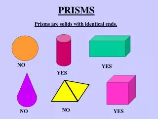
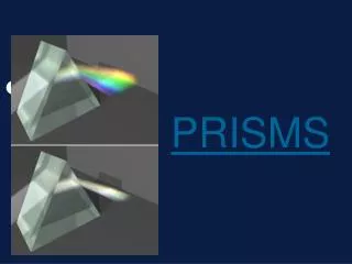
PRISMS. Prof: Vasudev Anand Rao. INTRODUCTION. A wedge of refracting medium Triangular cross-section with an apex and a base

PRISMS
E N D
Presentation Transcript
PRISMS Prof: Vasudev Anand Rao
INTRODUCTION A wedge of refracting medium Triangular cross-section with an apex and a base The angle α between the two surfaces is the refracting or apical angle of the prism. A line bisecting the angle is called the axis of the prism. The opposite surface is called the base. When prescribing prisms, the orientation is indicated by the position of the base ex: base-in , base-up
PRISM OPTICS • Light is refracted at each surface as it enters and exits the prism. • The ray is deviated towards the base of the prism. The net change in direction of the ray, angle D is called the angle of deviation • For a prism in air, the angle of deviation is determined by three factors: The refractive index The refracting angle α The angle of incidence
The image formed by a prism is • erect, • virtual and • displaced towards the apex of the prism.
NOTATION OF PRISMS The prism dioptre (Δ) • The power of a prism is said to be one diopter when it produces 1cm of linear apparent deviation of a light ray from where it would have otherwise traveled, measured 100 cm from the prism. • A 15 Δ prism produces 15 cm of deviation if a light ray when measured 1mt from the prism, the same prism would produce 7.5 cm of deviation measured 50 cm from the prism, and 30 cm of deviation when measured at 2mt from the prism.
USES: DIAGNOSTIC EVALUATION OF SUPPRESSION 4Δ BASE-OUT PRISM TEST • This test is used to detect suppression in small-angle esotropia and surgically corrected large angle squint. • When a 4Δ base-out prism is placed in front of a normal eye, the sudden displacement of the image to a parafoveal temporal point elicits a refixation movement. • No movement is seen in a microtropic eye, since the image is shifted within the central suppression scotoma. • Movements of the other eye also do not occur. 4Δ base-out prism test
DIAGNOSIS AND ACCURATE ASSESSMENT OF STRABISMICDEVIATIONS • The alternate cover test is performed first. • Prisms of increasing strength placed in front of one eye with the base opposite the direction of the deviation . ie base-in in a divergent strabismus. • Alternate cover test is performed. End point is reached when first neutralization and then reversal of the corrective occurs. • The angle of deviation then equals the strength of the prism. Prism bar cover test
KRIMSKY TEST WITH PRISM • Hirschberg’s test to locate the corneal reflex. • Placing the prism with appropriate base and gradually increasing the power in front of the fixing eye unless the corneal reflex is central – base out for esotropia, base in for exotropia. • The strength at which the corneal reflex is symmetrical is estimated.
MEASUREMENT OF AC/A (convergence ratio) • Denotes how many prism dioptres of convergence is produced for each dioptre of accommodation. The normal ratio is 3Δ-5Δ. • Important in the classification and management of accommodative and non-accommodative esotropia AC/A is calculated as: AC/A = (Δ¹- Δ²) / D , where • Δ¹ is the original deviation in prism dioptre, Δ² is the deviation in prism dioptre after putting the lenses and D is the power of new lens in dioptres
COMPONENTS OF DIAGNOSTIC OPHTHALMIC EQUIPMENT: The diagnostic instruments that contain prisms are: • Maddox double prism • Synaptophore • Direct ophthalmoscope • Indirect ophthalmoscope • Operating microscope • Slit lamp • Goldmann applanation tonometer • Stereoscopes • Pachymeter • Keratometer
PORRO-ABBE PRISM PORRO PRISM SLIT LAMP OPERATING MICROSCOPE
INDIRECT OPHTHALMOSCOPE STNAPTOPHORE
USES: THERAPEUTIC • To restore and maintain BSV in children with late onset strabismus, usually esotropia, who are awaiting surgical treatment. • To obtain BSV if there is a small surgical under-correction or overcorrection. Prism Exercises: useful when the angle of deviation is 15° or less, and patient can appreciate diplopia. • prism enabling the patient to fuse two images is held in front of one eye. • strength gradually decreased. • patient encouraged to fuse the images at whatever distance it is possible. • the object is then moved nearer and further, while patient tries to maintain fusion without assistance of prisms.
Prism vergence • To fuse diplopia: starting at the distance at which the diplopia is maximum, a prism bar is placed in front of one eye and the prism is gradually increased until the images join and fusion can be maintained without undue effort. • If this is impossible, fresnel prisms can be fitted as bifocals • To improve the cosmesis of a blind or disfigured eye or prosthesis. A base down prism can make hypotropic prosthesis appear slightly higher to the observer.
FRESNEL PRISMS: Composed of concentric annular rings. • Principle: the prism apex deviates light just as much as any other part of the lens. • Sheet of prism apices on a thin base sheet used to obtain a prismatic effect across the lens without creating additional lens thickness. • Method of choice for temporary use. Upto 30° can be applied to either eye but high powered prisms may not be tolerated.Upto 20° can be worn comfortably • The prism is normally fitted to the back surface of the spectacle lens.