Amebiasis (Amebic Dysentery): Symptoms, Diagnosis, and Treatment
Learn about the causal agent Entamoeba histolytica, morphology, life cycle, pathology, laboratory diagnosis, microscopy methods, and immunodiagnosis for Amebiasis. Differentiation from non-pathogenic species and molecular diagnostic methods are also discussed.

Amebiasis (Amebic Dysentery): Symptoms, Diagnosis, and Treatment
E N D
Presentation Transcript
Amebiasis (Amebic Dysentery) By: Assistant lecturer Ahmed Essam
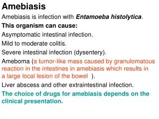
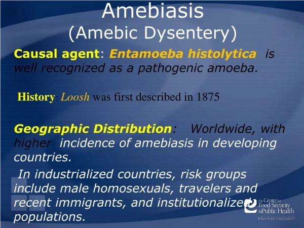
Amebiasis(Amebic Dysentery) Causal agent: Entamoeba histolytica is well recognized as a pathogenic amoeba.
Morphology • Different form of E. histolytica; • 1- trophozoite • 2- precyst • 3- cyst(1, 2, 4 nuclei)
Trophozoite chractere • Size: 12-60μm in diameter; • Non-invasive form / E. dispare • Invasive form contain RBC, E. histolytica • Pseudopodia: • Motility: Ectoplasm: • Endoplasm: may be contain ingested RBC invasive form Non-invasive form
Life cycle Life cycle
Laboratory Diagnosis • Entamoeba histolyticamust be differentiated from other intestinal protozoa including: E. coli, E. hartmanni, E. dispare,…… • Differentiation is possible, but not always easy, based on morphologic characteristics of the cysts and trophozoites. • The nonpathogenicEntamoeba dispar, however, is morphologically identical toE. histolytica, and differentiation must be based onisoenzymatic or immunologic analysis. • Molecular methods are also useful in distinguishing between E. histolytica and E. dispar.
Microscopy • Microscopic identification This can be accomplished using: • Fresh stool: wet mounts and permanently stained preparations (e.g., trichrome). • Concentrates from fresh stool: wet mounts, with or without iodine stain, and permanently stained preparations (e.g., trichrome).
Trophozoites ofEntamoeba histolytica(trichrome stain) A B Microscopy Each trophozoite has a single nucleus, which has a centrally placed karyosome and uniformly distributed peripheral chromatin.
Trophozoites ofEntamoeba histolyticawith ingested erythrocytes (trichrome stain) The ingested erythrocytes appear as dark inclusions. Erythrophagocytosis is the only morphologic characteristic that can be used to differentiateE. histolyticafrom the nonpathogenicE. dispar.
Cysts ofEntamoeba histolytica /E. dispar • GHI Cysts ofEntamoeba histolytica, permanent preparations stained with trichrome.
Immunodiagnosis(Antibody Detection) • 1- Antibody detection • 2- Antigen detection may be useful as an adjunct to microscopic diagnosis • The indirect hemagglutination(IHA) • The EIA test detects antibody specific for E. histolytica.
Molecular diagnosis • In reference diagnosis laboratories, PCR is the method of choice for discriminating the pathogenic species(E. histolytica).
Entamoeba coli is a non-pathogenic species of Entamoeba that frequently exists as a commensalparasite in the human gastrointestinal tract. E. coli (not to be confused with the bacteriumEscherichia coli) is important in medicine because it can be confused during microscopic examination of stained stool specimens with the pathogenicEntamoeba histolytica
Entamoeba coli 6-8 nuclei