Download
1 / 23
240 likes | 556 Vues
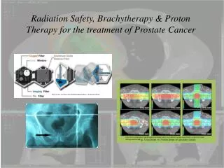
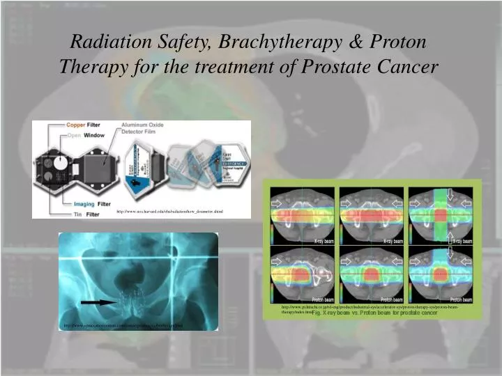
Radiation Safety, Brachytherapy & Proton Therapy for the treatment of Prostate Cancer. http://www.pi.hitachi.co.jp/rd-eng/product/industrial-sys/accelerator-sys/proton-therapy-sys/proton-beam-therapy/index.html. http://www.upmccancercenters.com/cancer/prostate/radbratherapy.html.

E N D
Radiation Safety, Brachytherapy & Proton Therapy for the treatment of Prostate Cancer http://www.pi.hitachi.co.jp/rd-eng/product/industrial-sys/accelerator-sys/proton-therapy-sys/proton-beam-therapy/index.html http://www.upmccancercenters.com/cancer/prostate/radbratherapy.html http://www.uos.harvard.edu/ehs/radiation/how_dosimeter.shtml
Radiation Safety & Radiation Therapy - Outline • Dosimetry is the measurement of radiation dose. • Dosimetry tracks exposure and monitors individual external radiation exposures. • Dosimetry use ensures that we are following the principle of ALARA, keeping exposures As Low As Reasonably Achievable. • One job of a medical physicist is that of radiation safety officer and chief dosimetrist keeping the medical staff and patients safe. • Discuss safety and then investigate proton therapy and learn how it works. • We’ll contrast proton therapy for prostate cancer treatment with x-ray therapy and brachytherapy.
Radiation Safety - Film Dosimeters • Film dosimeters, or film badges, consist of layered components. • Imagine a sandwich with the following layers starting from the top: • - the badge front, with a window for exposure; • - filters that selectively filter out certain types of radiation; • - films to detect the radiation; • - more filters perhaps; • - then the badge cover and clip to attach the dosimeter to the individual’s clothing. • After a designated period of exposure, the film is taken out of the “sandwich” badge, developed, and • read on a densitometer, which reads the amount of darkening on the film. • The amount of darkening is proportional to the radiation exposure.
Radiation Safety - Film Dosimeters • The Luxel body badge contains a sheet of radiation-sensitive aluminum oxide sealed in a light and moisture proof packet. When atoms in the aluminum oxide sheet are exposed to radiation, electrons are trapped in an excited state until irradiated • with a specific wavelength of laser light. The released energy of excitation, which is given off as visible light, is measured to determine radiation dose. • The packet contains a series of filters designed so that the energy and type of radiation can be determined. • In order for the radiation type and energy to be determined, the dosimeter must be worn so that the front of the dosimeter faces towards the source of radiation. Luxel • Body dosimeters are among the most sensitive dosimeters available. The minimum detectable dose is 10 mGy (1 millirem) for x-rays and gamma rays and 0.1mGy (10 millirem) for energetic beta radiation. http://www.uos.harvard.edu/ehs/radiation/how_dosimeter.shtml
Radiation Safety - Film Dosimeters • Advantages • The dose measurements for various film badges range between 0.1mGy – 15mGy (10 mrem to 1500 mrem) for gamma and x-radiation, and 0.5mGy – 10mGy (50 mrem to 1000 rem) for beta radiation. • Film badges can distinguish between penetrating radiation (high, medium, and low photon energies) and non-penetrating radiation (beta and x-ray radiation less than 20 keV). • Film dosimeters are practical because they are small, lightweight, and relatively inexpensive. • Disadvantages • The response of the film to radiation is energy dependent; at energies less than 300 keV, the response tends to increase. • The films cannot be read immediately and provide no radiation protection. • Environmental conditions such as heat and humidity will affect the film’s response to radiation. • Film badges may be left or lost at the site of the radiation accident • They may be contaminated with radioactive materials, which will lead to a false higher result.
Radiation Therapy with Protons • Has been around since the 1950’s in limited forms and proton beams offer the potential for improved distribution of radiation dose to tumors than traditional techniques • of using x- or gamma-rays or electron beams. • The improvement is due to the Bragg peak of a proton beam and the deposition of proton beam energy at the end of the range rather than along the entire trajectory. • Protons slow down relatively fast when entering biological tissue, loosing energy in atomic or nuclear interactions events. This reduces the energy of the protons, which in turn causes increased interaction with orbiting electrons energy. Maximum interaction with electrons occurs at the end of range causing maximum energy release within the targeted area with very little scatter. • The depth at which the peak occurs can be controlled by the amount of energy the protons are given by their accelerator. • The proton's dose of radiation is released in an exact shape and depth within the body. Tissues in front of the target receive a very small dose, while tissues adjacent to the tumor receive virtually none. • Proton beam therapy has demonstrated success for the treatment of selected tumors. More than 20,000 patients have been treated with protons or light ions in research laboratories or hospitals around the world and it costs an average of $50,000 to treat prostate cancer with protons...twice as much as with x-rays.
Radiation Therapy with Protons • Experimentally the range of a 125 MeV proton in tissue is 12 cm, while that of a 200 MeV proton is 27 cm. • It is clear that protons with enough energy can penetrate to any part of the body. • The proton proceeds through the tissue in very nearly a straight line (very little scatter), and the tissue is ionized at the expense of the energy of the proton until the proton is stopped. • The dosage is proportional to the ionization per centimeter of path, or specific • ionization, and this varies almost inversely with the energy of the proton. • Thus the specific ionization or dose is many times less where the proton enters the tissue at high energy than it is in the last centimeter of the path where the • proton is brought to rest. • Besides this very precise energy loss, the relative biological effect for protons is far more important than for photons. • Protons are much more ionizing than x- or gamma ray photons.
Radiation Therapy with Protons • The Bragg peak for electrons, protons and photons. • By adjusting the energy of the protons we can control the depth at • which they deposit their energy. • Protons are produced in accelerators and the accelerator ultimately • determines their energy. • The protons are steered from accelerator to patient by • using large magnetic fields. http://www.oncoprof.net/Generale2000/g08_Radiotherapie/Images/PicBragg.gif http://dd.dynamicdiagrams.com/wp-content/uploads/2007/12/proton2.jpg
Radiation Therapy with Protons • One of the gantries at the Northeast Proton Therapy Center – a joint venture between Harvard University and Massachusetts General Hospital. • The left picture shows the gantry structure during construction with the steel assembly being visible. • The right picture shows the gantry treatment room during treatment. • The beam delivery nozzle is able to rotate 360 degrees around the movable patient couch. http://www.aapm.org/meetings/05AM/pdf/18-4016-65735-22.pdf http://www.aapm.org/meetings/05AM/pdf/18-4016-65735-22.pdf
Radiation Therapy with Protons • The accelerator, which is housed in a 25,000-square-foot facility, funnels protons into a 40-foot- wide circular track known as a cyclotron. The cyclotron speeds up protons to higher energy levels. • The patient enters one of three chambers, depending on the type of treatment, and lies on a gurney- like bed. A computer-controlled proton-firing nozzle positions itself over the target area. • Meanwhile, magnets guide a beam of protons along the center of a long, narrow tube. The beam races toward a gantry, which rotates around the patient as its nozzle fires protons at the tumor. • Accelerator facility cost ~ $100 Million http://www.popsci.com/files/imagecache/article_image_large/articles/tumor_485.jpg
Radiation Therapy with Protons • Depth dose curves for protons in water as a function of the incident proton energy • As the protons energy increases they penetrate farther into the target material. • By knowing the depth of the tumor you can control the dosage delivered by selecting a proton of sufficient energy to reach the treatment spot. http://www.rptc.de/fileadmin/user_upload/rptc/Jahresberichte/JB_2012/englisch/Bilder/Fig.%2010.JPG
Radiation Therapy with Protons • You can use proton beams with different energies to cover the tumor by depositing the energy at various depths in the tumor. • This is called the Spread Out Bragg Peak, or SOBP. • Here you can deposit a homogenous dose in the target region using only a single proton beam direction • Thus you can provide an effective treatment to irradiate the entire tumor. • However, in practice usually multiple proton beams are used all incident from different directions. http://sunse.jinr.ru/~molod/concept/pr_ph_depth.gif http://bjr.birjournals.org/content/79/937/24/F1.large.jpg
Radiation Therapy with Protons • Irregularly shaped lesions located near critical structures, tumors in children, and large tumors near any critical organ are well suited for proton beam therapy. • Protons have a physical advantage over gamma rays and x-rays when it comes to sparing normal tissues. • Protons deposit most of their radiation energy in what is known as the Bragg Peak, which occurs at the point of greatest penetration of the protons in tissue. • The exact depth to which protons penetrate, and at which the Bragg Peak occurs, is dependent on the energy or modulation of the proton beam. • This energy can be very precisely controlled to place the Bragg Peak within a tumor or other tissues that are targeted to receive the radiation dose. • Because the protons are absorbed at this point, normal tissues beyond the target receive very little or no radiation. • Proton energy can be adjusted to match the depth of the target with a sharp drop in dose beyond the Bragg Peak.
Radiation Therapy with Protons • Filter block used to select out various proton energies. http://www.aapm.org/meetings/05AM/pdf/18-4016-65735-22.pdf
Radiation Therapy with Protons • Tumors can have very irregular shapes and can be located close to critical organs. • Every patient’s tumor shape, size and location are unique. • Patient specific hardware, which helps sculpt the proton beam, is customized to maximize the dose to the tumor while minimizing the dose to normal structures. • The shaping of the proton beam can also be controlled by magnetically scanning across the tumor volume. • Aiming proton beams, each with customized field shaping, from various directions further ensures that the dose to normal tissues is reduced as much as possible, therefore reducing the risk of treatment related complications. • This is a mask that is used to treat a certain • geometry of tumor. The plate on the left • stops the protons from entering the patient • while the “hole” controls the shape of the • beam. http://www.massgeneral.org/radiationoncology/assets/ProtonTreatment/principlesProtonTh_1.jpg
Radiation Therapy with Protons • A significant proportion of patients treated in radiation oncology centers have prostate cancer. • Side effects of treatment generally include gastrointestinal (GI) and genitourinary (GU) damage due to photon therapies. • Large numbers of patients experience urinary frequency and diarrhea during treatment, and long-term, may suffer impotence, • incontinence, rectal fibrosis and bleeding, and extensive bowel fibrosis. • These side effects may cause a reduction in the quality of life. • Proton therapy may be able to deliver equivalent, doses to the prostate while sparing more • normal tissues when compared with photon based therapy. http://www.oncolink.org/treatment/article.cfm?c=9&s=131&id=425
Prostate Cancer - Prostate function • The prostate is a small walnut shaped gland that wraps around a tube called the urethra which carries urine and • semen out of the penis. • The prostate produces the white fluid found in semen, or the white fluid that contains sperm. • Prostate cancer is usually diagnosed with a blood test measuring the amount of prostate specific antigens • (PSA) in the body or by a physician conducting a rectal exam and manually examining the prostate or can be diagnosed using a PET scan. • Symptoms can include: • - Changes in urinary flow: frequency, urgency, hesitancy • - Frequent night time urination • - Painful urination • - Blood in urine http://www.igrt.com/prostate_cancer.asp
Prostate Cancer - Case Study An 82 year old male with prostate cancer diagnosed 17 years ago, status post TURP (Transurethral Resection of the Prostate), radiation therapy, orchiectomy in 1995. He has recent rising PSA. Bladder prostate http://www.medscape.com/viewarticle/549296_2 http://www.petscaninfo.com/zportal/portals/phys/clinical/petct_case_studies/prostate/prostate_case4/prostate_case4.swf • PET scan using 11.1mCi 18F FDG • administered left arm IV with CT and fused CT/PET scan • showing possible recurrence of prostate cancer. • Normal MRI image of the lower abdomen showing the bladder and prostate.
Prostate Cancer - Brachytherapy treatment • Prostate Brachytherapy, also known as a seed implantation, is often done in the • operating room. It delivers a very high dose of radiation to your tumor by inserting radioactive seeds directly into your prostate gland under ultrasound guidance while you are asleep. • Iodine or palladium are most commonly used. The seeds are about four millimeters • long and less than a millimeter in diameter. Sometimes both prostate brachytherapy and external radiation may be used to combat your tumor. http://www.igrt.com/prostate_cancer.asp# http://www.upmccancercenters.com/cancer/prostate/radbratherapy.html • Depending on the stage of your disease, you often have more than one treatment option to consider. Several factors should be taken into • account when choosing these options, including potential benefits and risks.
Prostate Cancer - Brachytherapy treatment • Advantages of Brachytherapy • Unlike major surgery or daily radiation treatments, brachytherapy causes little interruption in your daily activities. • In addition, this treatment usually preserves continence and causes erectile dysfunction less frequently than surgery or external beam radiation therapy. • Disadvantages of Brachytherapy • Disadvantages, such as infection and bleeding, are those of a 90-minute surgical procedure. • Death is a risk of all surgery involving general anesthesia, but is an extremely rare occurrence with this procedure.
Prostate Cancer - Brachytherapy treatment • The following side effects are generally caused by the radiation emitted by the seeds in the prostate. The effects may last for two to 12 months after the implant and will decrease gradually as the seeds lose their radioactivity. • Frequent urination, burning with urination and urinary urgency occur in 75 percent of men, six weeks to three months after seed implant. These side effects generally • last for a few weeks. Dietary changes and bladder medications can control symptoms. • Urinary obstruction occurs occasionally, due to an initial swelling of the prostate caused by the seeds. Obstruction is a higher risk for patients who had • obstructive symptoms prior to surgery. • Diarrhea or a change in bowel habits occurs very rarely. • Erectile dysfunction, with brachytherapy as the sole treatment, occurs when radiation thickens the walls of blood vessels, limiting the blood supply to the nerves responsible for erections. It is seen in 30-35 percent of men five years after • seed implantation. Vascular problems caused by smoking, arteriosclerosis, or diabetes can significantly increase the chances of erectile dysfunction after radiation.
Prostate Cancer - Proton Therapy • The six images compare the dose distribution of x-ray beams with proton beams. • A tumor of the prostate gland appears in each MRI/CT image. The various colors indicate the intensity of the dose deposited in the tissue. Red is the maximum dose, followed by orange, yellow, green, blue, and purple, the minimum. • The two images on the left compare the dose distribution of a single X-ray beam and • a single proton beam. • The four images on the right show the effect of multiple beams. Even with multiple X- • ray beams (top right) there is still substantial dose to healthy tissue. • However, with the two lateral proton beams (lower middle), the dose nicely • conforms to the shape of the tumor. http://www.pi.hitachi.co.jp/rd-eng/product/industrial-sys/accelerator-sys/proton-therapy-sys/proton-beam-therapy/index.html • Regardless of the quantity of x-ray beams used, there will be two to three times more integral dosage to normal tissue than with protons.
Prostate Cancer - Proton Therapy • Advantages • With these charged particles physicians can precisely focus the destructive characteristics of a proton beam in the target volume and greatly reduce the damage given to the normal cells and tissues. • This contrasts with X-rays, which are electromagnetic radiation in the high energy or high frequency portion of the electromagnetic spectrum, with very little • three-dimensional controllability resulting in greatly reduced ability to avoid unwanted damage to patient's normal tissues. • Damage to normal tissues is the cause of patient morbidity in all forms of therapy. • Disadvantages • Large size and costs of an accelerator and of the beam lines needed for the transport of the beam which coupled with medical and technical staff are passed on to patient. • Large technical staff to keep accelerator running.