Understanding Microscope Parts and Calculations for Effective Use
Explore the essential parts of a microscope, including its stage, objective lenses, and light source, to enhance your viewing experience. Learn how to calculate magnification using the ocular and objective lenses, and discover how to determine the actual size of specimens. This guide also covers key rules for creating biological diagrams, ensuring accurate and labeled illustrations. Ideal for students and enthusiasts, you'll gain valuable skills in microscopy and biological drawing techniques.

Understanding Microscope Parts and Calculations for Effective Use
E N D
Presentation Transcript
MICROSCOPE LAB WHAT DO YOU SEE? WHAT CAN YOU SEE?
Parts of the MicroscopeFill and Chill • http://www.biologycorner.com/microquiz/
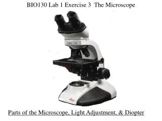
PARTS OF THE MICROSCOPE Stage: Supports the slide. The central opening in the stage allows light to pass through. Stage Clips:Keep the slide in position Diaphragm: Regulates the amount of light reaching the object being viewed. Ocular Lens: Viewing (10X)/Eyepiece Coarse Adjustment knob: Moves the body tube up or down so you can get the specimen in focus. It is used with the low power objective only! Fine Adjustment Knob:Used with med or high power to get the specimen in sharper focus Arm:Part of the microscope you carry the microscope with Base:Supports the microscope Light Source:Provides light making it easier to view the specimen Objective:Low (4X), Medium (10X) and High (40X)Attached to the revolving nosepiece Body Tube:The long tube that holds the eyepiece and connects it to the objective
CALCULATIONS 1. What is the MAGNIFICATION of the OCULAR LENS? The magnification of the ocular lens is ________X 2. Calculating the MAGNIFICATION of the MICROSCOPE. Magnification of microscope =ocular lens x objective lens Magnification of microscope (4X objective) ___ x ___ = _____ Magnification of microscope (10X objective) ___ x ___ = _____ Magnification of microscope (40X objective) ___ x ___ = _____
3. What is the ACTUAL SIZE of the SPECIMEN you looked at with the microscope? Actual size of specimen (mm) = Field of view (see table above) # of specimens to fit across field of view Magnification of MicroscopeField of view 40X (low) 4.2 mm 100X (medium) 1.72 mm 400X (high) 0.42 mm
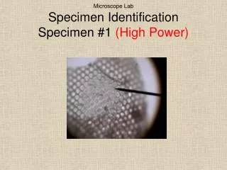
Example of Calculations for Science 10 Using the 4X objective of a compound light microscope, you view the picture at the side. What is the magnification of the microscope? What is the actual size of the specimen?
Rules for Biological Diagrams • Unlined paper and a sharp pencil • Leave an empty margin of about 1 cm all around your page • When drawing cells, choose only one cell (unless otherwise required) and show the edge of neighboring cells to show the connection • Draw outline of your subject with clear and unbroken lines. • Your drawing should be about ½ of the page • Use Figure # and name as your heading • Always indicate the magnification of the illustration below your drawing • Use stipples for darkened areas • Always label to the right with a ruler • Measurement to the left • Never cross lines • Do little or no erasing • Small letters, no writing
Using a clean sheet of paper, complete a biological drawing. • Submit before leaving. • Thanks