Microscope Basics: A Guide for Biology Students
210 likes | 328 Vues
Explore the world of microscopes and cells with this comprehensive guide for biology students. Learn hands-on techniques, microscope operation, and cell observation exercises. Get ready to dive into the fascinating microscopic world!

Microscope Basics: A Guide for Biology Students
E N D
Presentation Transcript
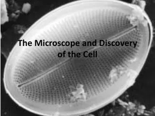
Lab 1The Microscope and the Cell BY 102 Zach Nolen
A few introductions • I’m a Master’s student in the Thacker lab • Studying sponge/cyanobacteria relationships • Teaching Portfolio
Now about you • Fill out your notecard with the following: • Name (you wish to be called) • Major • Reason taking class (be honest) • 1 thing your looking forward to in this class • 1 thing your not looking forward to in this class • What’s your favorite food
Tips for Success • Read ahead • Do study guides • Work on lab manual throughout • Keep good notes • Ask questions • Study with others
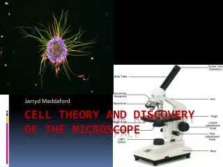
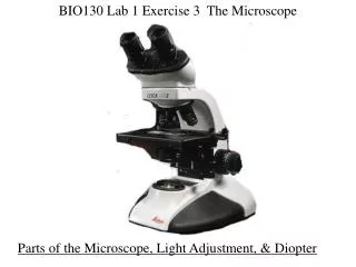
Microscope Introduction • Microscopes allow us to view very small objects • There are three main types of microscopes: compound light, dissecting, and electron microscopes. • For this class, we will be working with compound light microscopes.
Electron Microscopes Spider Pollen Diatoms
Exercise A: Use and Care of Microscope • Always carry scope with one hand on the arm and the other under the base. • Always clean scope when you are done. • Always start on the low objective (red), then work your way up in magnification. • Never use the coarse adjustment knob when using an objective higher than red. • If there are any issues with your scope, please let me know immediately.
Exercise B: Functions of the Microscope • Microscopes are very useful tools for biologists. • Not only can they be used to magnify things, they can also be used to measure things. • To calculate the total magnification of your scope, you simply multiply the ocular magnification x objective magnification • Take a few minutes to complete the chart in your lab manual.
Exercise C: Calibration • For this exercise, we will be using the letter “e” slides. • You will place the slide on the microscope with the “e” in the correct orientation, then view it through the microscope to observe how the scope transposes images.
Exercise D: Human Epithelial Cells • In this exercise, you will be preparing a wet mount of your own cheek cells. • Make sure that you are using a blank slide. • We will be doing a slightly different procedure than listed in the book. • After you have prepared your slide, use your microscope to find a cell on the red objective and let me check it. • After I check it, you will need to use the highest objective (blue) to find the cells for your drawing. • After you have finished your drawing, you can wash your slide off in the sink. You will be using the same slide for other exercises.
Exercise E: Plant Cells • For this exercise, you will be preparing a wet mount of an Elodea leaf to observe plant cells. • Elodea is a freshwater plant that is typically found in ponds. • Follow procedure in the book for slide prep. • You will draw this using the blue objective • After you finish your drawing, you will want to add a few drops of NaCl to the leaf and observe plasmolysis.
Exercise G: Demonstration of Osmosis • This exercise is demo that I will do to show the process of osmosis. • What is osmosis? • Osmosis is simply the movement of water molecules through a selectively permeable membrane from an area of higher concentration to an area of lower concentration.
Exercise H: Animal and Plant Cell Structures • This exercise is just a review of the various parts of plant and animal cells as well as their functions. • Figures 1.8 and 1.9 in your manual have labeled images of both. • You should be able to identify the different parts and also be able to give their function.
Review Questions • What are the 3 types of microscopes? • Which type will we use in lab? • What are 3 differences between animal and plant cells? • What is osmosis? • What is the purpose of mitochondria in the cell?
Before you leave lab! • Make sure you properly store your microscope • Clean up your work station • Wipe down your station • Turn in your microscope drawings
Before next lab! • Read Lab exercise 2: Nutrition and the Cell • Do the online prelab • Read the introduction in your manual (viii-xi) • Complete all questions in Lab 1 exercises that we covered, if not already done so