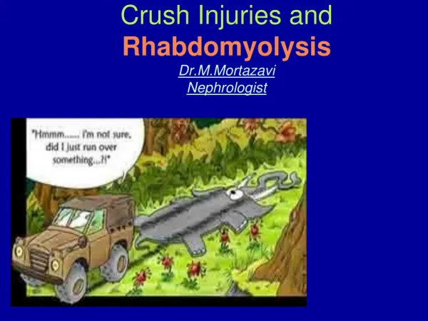
Download
1 / 12
140 likes | 709 Vues
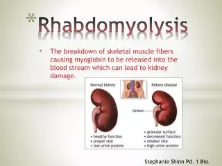
Rhabdomyolysis. in Equus caballus (horse) By R.Rampersadh (205515894). Possible human homologue (MIM number): 268200 MIA number: Phene ID 2032, Group 001158 Genes associated with Rhabdomyolysis in Equus caballus: GYS1: glycogen synthase 1 (muscle), also known as GSY, GYS.

E N D
Rhabdomyolysis in Equus caballus (horse) By R.Rampersadh (205515894)
Possible human homologue (MIM number):268200 • MIA number: Phene ID 2032, Group 001158 • Genes associated with Rhabdomyolysis in Equus caballus:GYS1: glycogen synthase 1 (muscle), also known as GSY, GYS. • Gene map location:2p21 • Symbol:PSSM • Cross Species Summary:Rhabdomyolysis. Disintegration of muscle fibres, with consequent excretion of myoglobin in the urine. • Summary:Valber et al. (1996) identified a polysaccharide storage myopathy (a glycogen storage disorder) with consequent excretion of myoglobin in the urine. associated with exertional rhabdomyolysis in Quarter-horse related breeds. • Species Specific Name:Equine rhabdomyolysis syndrome; Polysaccharide storage myopa
Inheritance:The disorder is familial, and there are suggestions of autosomal recessive inheritance. However, the published data are inconclusive on this point (Valberg et al., 1996). Dranchak et al. (2005) performed a segregation analysis that excluded all forms of single-locus inheritance other than autosomal dominant. • Single Locus:Yes • Characterised at a molecular level:Yes • Phenotype Considered a Defect:Yes • Clinial Features and Pathology:Widespread presence of subsarcolemmal vacuoles and PAS-positive inclusions (Valberg et al., 1996). • Human Genes and Disorders:MYOGLOBINURIA, ACUTE RECURRENT, AUTOSOMAL RECESSIVE. Recurrent myoglobinuria is characterized by recurrent attacks of rhabdomyolysis associated with muscle pain and weakness and followed by excretion of myoglobin in the urine.
CLINICAL SIGNS • Signs can be extremely variable and severely overlap with other muscle disorders. • In severe cases, they can be confused with signs of colic. • Adequate diagnosis requires examination of blood serum as well as muscle biopsy. Occasionally electromyography can be useful as well (Beech,1997). • Mild forms - muscle spasms and exhibits a slightly restricted gait, a transient stiffness, or shortened stride during exercise. • After exercise, if the episode persists - weight shifting, partial posturing or an anxious expression with muscle spasms. At times - violent and appear to mimic signs for colic. Profuse sweating may follow, along with limb weakness.
CLINICAL SIGNS CONT… • In severe cases - collapse and are unable to rise, coffee-colored urine (myogloburia) • In chronic cases - a horse will tie up under moderate exercise one day and not another day under more strenuous exercise. • In endurance horses - conditions of extreme physical exertion. Because these horses predominantly use Type I fibers -including rapid heart rate, dehyration, hyperthermia, synchronous diaphragmatic flutter and collapse.
DIAGNOSIS • - Exhibition of clinical signs - Elevated CK and AST concentrations - Muscle biopsies- Electromyography • MUSCLE BIOPSY SAMPLES- Muscles used most frequently for biopsies to diagnosis ER are the mid gluteal and semimembranous muscles, which are normally mostly comprised of Type II fibers. - Biopsies continually reveal that Type II fibers are more severely affected than Type I. In vitro testing has shown that thresholds of Ca++-induced calcium release in heavy sarcoplasmic reticulum fractions was lower in affected horses (Beech,1993). - Gluteal muscle stained with periodic acid-Schiff stain (PAS) helps to specifically diagnosis PSSM. In normal horses, the stain uniformly takes over the muscle completely throughout. In PAS-positive stains, only some fibers become stained, with noticable light areas around them (Valberg,1997).
DIAGNOSIS CONT… • ELECTROMYOGRAPHY (EMG)The technique uses an instrument called an electromyograph in order to record the physiological properties of muscle at rest. While this techinique is not widely used to diagnosis ER, it has proved to be helpful in detecting abnormal electrical activity in muscles which are sometimes produced by myotonic discharges (Beech,1997).
POLYSACCHARIDE STORAGE MYOPATHY • has been marked as a distinct cause of exertional rhabdomyolysis in certain breed lineages. • classified as a glycogen-storage disease characterized by high muscle glycogen concentration (1.5 to 4 times greater than those reported for horses with RER), accumulation of an abnormal polysaccharide, subsarcolemmal vacuoles, and fiber necrosis. • PSSM horses generally have a calm disposition and exhibit such signs as tucked-up abdomen, muscle fasiculations in the flank, stiff gait, camped-out stance and severe cramping of hindlimb and tricep muscles soon after beginning light exercise. • The accompaniment of high glycogen concentration with rhabdomyolysis has occurred in humans and other species as well. These are commonly due to heritable defects in genes coding for enzymes important in glycolysis in the muscle cell. It has become generally accepted as agenetic disorder • A diagnosis of PSSM is based on the presence of muscle fibers with subsarcolemmal vacuoles, dark periodic acid-Schiff (PAS) staining for glycogen, and most notably, amylase-resistant abnormal complex polysaccharide accumulation.
RECURRENT EXERTIONAL RHABDOMYOLYSIS (RER) • Due to abnormal regulation of intracellular calcium in skeletal muscles. It appears there is intermittent disruption of muscle contraction, particularly when horses susceptible to the condition are fit and have a nervous temperament. • Determination of the cause of chronic tying-up include a serum chemistry panel, blood vitamin E and selenium concentrations, urinalysis to determine electrolyte balance, dietary analysis, exercise testing, and muscle biopsy. • An exercise challenge test is useful to detect subclinical cases. In addition, quantifying the extent of exertional rhabdomyolysis during mild exercise is helpful in deciding how rapidly to reinstate training. • Diagnosis of recurrent exertional rhabdomyolysis is based on history, clinical signs, elevations in serum CK and AST, and muscle biopsy. Quarter Horses with PSSM have enhanced sensitivity to insulin, resulting in high muscle glycogen concentrations.
CONTROL • Thus, the ideal diet – provide >15% of digestible energy as fat and limits starch to <10% of daily digestible energy by limiting grain or replacing it with a fat supplement. • Caloric needs should be assessed first to prevent horses becoming obese on a high-fat diet. Improvement in signs of exertional rhabdomyolysis for horses with PSSM requires both dietary changes and gradual increases in the amount of daily exercise and turn-out. • Management of recurrent exertional rhabdomyolysis - decreasing the triggering factors for excitement and pharmacologic alteration of intracellular calcium flux with contraction. • Management changes that may decrease excitement include minimizing stall confinement by using turn-out or a hot walker, exercising and feeding horses with recurrent exertional rhabdomyolysis before other horses, providing compatible equine company, and the judicious use of low-dose tranquilizers during training.
CONTROL CONT… • A high-fat, low-starch diet – decreases excitement. In contrast to PSSM, horses that have RER often require higher caloric intakes. At these high caloric intakes, specialized feeds designed for exertional rhabdomyolysis are necessary, as additional vegetable oil or rice bran cannot supply enough calories for athletes in intense training. Hay should be fed at 1.5-2% of body weight and high-fat, low-starch concentrates should be selected that provide ≤ 20% of daily digestible energy as nonstructural carbohydrate and 20-25% of digestible energy as fat. • Dantrolene given 1 hr before exercise to horses that are not fed prior to exercise may decrease the release of calcium from the calcium release channel. • Phenytoin has also been advocated as a treatment. Therapeutic levels vary, so oral dosages are adjusted.