Download
1 / 49
510 likes | 2.13k Vues
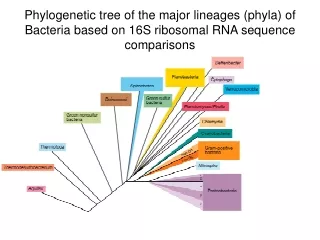
3. Phylum Proteobacteria. GRAM NEGATIVE BACTERIA Wide morphological and metabolic diversity . Clinical , environmental , and industrial relevance Phylum with highest number of cultured representatives 5 groups ( based on 16S rRNA ): a , b , g , d, and e.

E N D
3. Phylum Proteobacteria GRAM NEGATIVE BACTERIA Widemorphological and metabolicdiversity. Clinical, environmental, and industrial relevance Phylumwithhighestnumber of culturedrepresentatives 5 groups(basedon 16S rRNA): a, b, g, d, ande
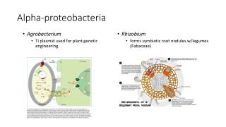
3. Phylum Proteobacteria 3.1. a-Proteobacteria: Rhizobium N2 fixing organisms: Some cyanobacteria (Bacteria) Some anoxygenic phototrophs (Bacteria) Some chemolithotrophs (Bacteria/Archaea) Some chemoorganotrophs (Bacteria/Archaea) Some chemoorganotrophs (Bacteria) FREE LIVING SYMBIONTS E.g. symbionts of leguminous plants
3. Phylum Proteobacteria 3.1. a-Proteobacteria: Rhizobium RECOGNITION NOD FACTORS
3. Phylum Proteobacteria 3.1. a-Proteobacteria: Rhizobium
3. Phylum Proteobacteria 3.1. a-Proteobacteria. Agrobacterium pTi
3. Phylum Proteobacteria 3.1. a-Proteobacteria: “acetic acid bacteria” They oxidize sugars and alcohols Acidophilic ETHANOL ETHANAL Gluconobacter ACETIC ACID e- e- Acetobacter H20 O2
3. Phylum Proteobacteria 3.1. a-Proteobacteria: Caulobacter Prosthecae: cytoplasmic extrusions (stalks, hyphae, or appendages) Budding division (mother cells retain their original identity) Attachment, increased surface-to-volume ratio, reduced cell sinking Aquatic bacteria (either planctonic or benthonic) PROSTHECAE
3. Phylum Proteobacteria 3.2. b-Proteobacteria. Neisseria Aerobic diplococcus Penicillin sensitive Pathogens and normal microbiota Carrier state N. meningitidis (“meningococcus”) Aerosols Nasopharynx Blood stream: Meningitis (sudden onset of headheache, vomiting and stiff neck) Intravascular coagulation shock
3. Phylum Proteobacteria 3.2. b-Proteobacteria: Neisseria N. gonorrhoeae (“gonococcus”) - Very sensitive to environmental stress (drying, sunlight, UV light…) - High incidence (STD, ETS) - Mild symptoms* in women (asymptomatic carriers). PID - Complications if untreated Sexual contact Mucous membranes of the genitourinary tract Reasons?
3. Phylum Proteobacteria 3.2. b-Proteobacteria. Acidithiobacillus Chemolithotroph: donor: Fe2+ / acceptor: O2 They use large amounts of substrate (they fix CO2) pH acidic (acidiphilic) Acid mine drainage (formation of H2SO4 and Fe3+) (O2, water and bacteria) Biolixiviation Thiobacillus ferrooxidans
3. Phylum Proteobacteria 3.2. b-Proteobacterias. Zooglea WASTEWATER TREATMENT BOD: Biological Oxygen Demand
3. Phylum Proteobacteria 3.3. g-Proteobacteria: Legionella Rods Complex nutritional requirements (Fe) Resistent to b-lactamic Terrestrial and aquatic habitats Waterborne, aerosols (no person-to-person) Pontiac fever Pneumonia LEGIONELLOSIS L. pneumophila Emerging disease (1976) Cases per 100.000 inhabitants. Spain
3. Phylum Proteobacteria 3.3. g-Proteobacteria: Legionella Legionella inside an alveolar macrophage
3. Phylum Proteobacteria 3.3. g-Proteobacteria: Haemophilus Haemophilus influenzae
3. Phylum Proteobacteria 3.3. g-Proteobacteria: Pseudomonas • More than 100 species • Some species utilize over 100 different compounds • Aerobic chemoorganotrophic rods • Some chemolitotroph (H2, CO) • Some anaerobic (NO3- / fermentation) • Sugar oxidation: Entner-Doudoroff pathway • Fluorescent pigments
3. Phylum Proteobacteria 3.3. g-Proteobacteria: Pseudomonas Pseudomonas aeruginosa Opportunisticpathogen; intrahospitalaryinfectionsResistenceplasmids (plasmids R) Respiratory tract infections Skin infections Bacteremia Endocarditis Joint infections Fastrointestinalinfections UTI Eye infections CNS infections Upper respiratory tract infections Cystic fibrosis complications
3. Phylum Proteobacteria DEGRADATIVE MEGAPLASMIDS 3.3. g-Proteobacteria: Pseudomonas
3. Phylum Proteobacteria 3.3. g-Proteobacteria: Family Enterobacteriaceae Enteric bacteria • Facultatively aerobic, gram negative rods • Fermentation of sugars to organic acids • Some respire NO3- to NO2- (never to N2) Virulence factors and pathogenicity
3. Phylum Proteobacteria 3.3. g-Proteobacteria: Family Enterobacteriaceae Escherichia Warm blooded animals Gut microbiota (comensals) Pathogenic strains: E. colienterotoxigenic (ECET) E. colienteropathogenic (ECEP) E. colienterohemorrhagic (ECEH) (STEC) E. coli O157:H7 E. coli O104:H14 Gastroenteritis Hemolytic uremic syndrom (HUS) Urinary tract infections (UTI)
3. Phylum Proteobacteria 3.3. g-Proteobacterias. Familia Enterobacteriaceae Salmonella Habitat: gastrointestinal tract of wild and domesticanimals, birds, pets, and insects. Zoonosis. Mainpathogenicserovars: S. typhimurium: salmonellosis (enterocolitis) S. typhi: typhoidfever
3. Phylum Proteobacteria 3.3. g-Proteobacteria: Family Enterobacteriaceae Shigella Reservoir: infectedgut S. dysenteriae: - Shigellosisorbacillarydysentery (“disentería bacilar”) - HUS Shigella cells invading intestinal epithelium
3. Phylum Proteobacteria 3.3. g-Proteobacteria: Family Enterobacteriaceae Yersinia: Y. pestis PLAGUE (bubonic, pneumonic and speticemic). Zoonotic disease. Gangrene and black spots (“black death”) Buboes (“bubones”) Bacteria in lung tissue
3. Phylum Proteobacteria 3.3. g-Proteobacterias: Vibrio Curved and straight rods Aquatic media Many species, some pathogenic: V. cholerae: cholera CHOLERA TOXIN
3. Phylum Proteobacteria 3.3. g-Proteobacteria: Photobacterium Autoinduction: “quorum sensing” FMN luciferase FMNH2 + O2 + RCHO FMN + RCOOH + H2O + luz NADH Inducer molecule: acyl homoserine lactone (AHL) NAD+
3. Phylum Proteobacteria 3.4. d-Proteobacteria: Bdellovibrio “Predator” of other bacteria
3. Phylum Proteobacteria 3.4. d-Proteobacteria: SULFATE (AND SULFUR) REDUCING BACTERIA (SRB)* Desulfo- (generally) or Desulfuro- They reduce sulfate/sulfur(acceptors) Final products: H2S Strict anaerobes (anoxic environments) Group I: Acetate cannot be used as donor Sulfate reducing bacteria Group II: H2 and acetate can be used as donors Sulfate reducing bacteria Fixation of CO2 (acetil-CoA pathway) Group III: H2 or organic matter as donors S and sulfite reducing bacteria (NEVER sulfate) Donors H2 O. M. *Some sulfate-reducing bacteria are not delta-proteobacteria
3. Phylum Proteobacteria 3.4. d-Proteobacteria: SULFUR AND SULFATE REDUCING BACTERIA Marine sediments m.o. O2 +0.8 NO3- Mn+4 Fe+3 0 E’ (V) Consortia of SRB/methane oxidizing Archaea SO4-2 Stratification of electronic acceptors Limited organic matter Most important metabolism: sulfate reduction Competence (or not---) with methanogenic Archaea -0.2 CO2
3. Phylum Proteobacteria 3.4. d-Proteobacteria: MYXOBACTERIA Vegetative cells: long rods without flagella (names: Myxo…). Gliding. Fruiting bodies: cell-to-cell communication and differentiation
3. Phylum Proteobacteria 3.5. e-Proteobacteria: Campylobacter and Helicobacter (both microaerophilic) Campylobacter: acute gastroenteritis, food borne disease (chicken meat…)
Helicobacter: gastritis and ulcers http://www.nobelprize.org/nobel_prizes/medicine/laureates/2005/marshall-lecture.pdf
Nitrospira (independent phylum) Nitrifying Archaea 3. Phylum Proteobacteria 3.6. OTHER PROTEOBACTERIA: NITRIFYING BACTERIA a, b, g , and d Proteobacteria Electron donors: NH4+ and NO2- / Acceptor: O2 (Nitroso…/ Nitro…) They consume large amounts of substrate: chemolithoautotrops/ chemolithoheterotrophs Soils and waters; ammonia-rich sites; leaching of NO3- Nitrification NH4+ NO2- NO3- Nitrosomonas Nitrobacter AMO: ammonia monooxygenase NOR: nitrite oxidoreductase
3. Phylum Proteobacteria 3.6. OTHER PROTEOBACTERIA: SULFUR OXIDIZING BACTERIA a, b, g, eProteobacteria Donors: H2S, S0, S2O32-, metallicsuflides, H2 / Acceptor: O2 (sometimes NO3- ) S0accumulationinsideoroutsidethecell Acidithiobacillus: acidiphilic (A. ferrooxidans FeS2, Fe2+); autotrophs Thiomargarita: anaerobicoxidation (acceptor: NO3-)
3. Phylum Proteobacteria 3.6. OTHER PROTEOBACTERIA: PURPLE BACTERIA Anoxygenic phototrophs Reaction centre: bacteriochlorophylls Antenna pigments: BChl + carotenoids Photosynthetic systems: invaginations of the cytoplasmatic membrane
e- e- e- 3. Phylum Proteobacteria 3.6. OTHER PROTEOBACTERIA: PURPLE BACTERIA ATP: Cyclic photophosphorilation NAD(P)H: Reverse electronic flow NAD(P)+ NAD(P)H BChl EXCITED BChl EXCITED Eo (-) cte- cte- H+ H+ H+ H+ H2S S0 Fe2+ S0 SO42- Fe3+ BChl BASAL BChl BASAL Eo (+) LIGHT NO external electron donors for ATP External electron donors for NAD(P)H Imagesproperty of Fernando Santos
3. Phylum Proteobacteria 3.6. OTHER PROTEOBACTERIA: PURPLE BACTERIA g-Proteobacteria (“purple sulfur bacteria”): highly sensitive to O2; highly tolerant to [H2S] S compounds as electron donors. Photoautotrophs (Calvin cycle) Photoheterotrophs N2 fixers a/b-Proteobacteria (“purple nonsulfur bacteria”): more tolerance to O2; very sensitive to high [H2S] Chenoorganotrophs, Photoheterotrophs, photoautotrophs N2 fixers Anoxic areas blooms Sulfide-rich waters (SRB) Meromictic lakes, microbial mats
Microbialmats: stratificationof microbialpopulationsdrivenbyenvironmentalgradients Imagesproperty of Fernando Santos
3. Phylum Proteobacteria 3.6. OTHER PROTEOBACTERIA: METHANOTROPHS Methylotrophs vs. methanotrophs Biotic and abiotic methane Methanotrophs vs. methanogens Where does the methane come from?
3. Phylum Proteobacteria 3.6. OTHER PROTEOBACTERIA: METHANOTROPHS (Methylo…) Type I Type II
Methane as energy source Methano Methanol Formalhehyde CO2 Methane monooxygenase
Methane as carbon source Methane Methanol Formalhehyde biomass Type I Type II Ribulose monophosphate pathway Serine pathway
3. Phylum Proteobacteria 3.6. OTHER PROTEOBACTERIA: RICKETTSIAS a/g Proteobacteria Obligate intracellular parasites* Induce phagocytosis, do not survive outside hosts Highly specific energy metabolism*, synthesize few compounds Transmitted by arthropod vectors Damage to blood vessels Epidemic tifus (Rickettsia prowazekii) Vector: human louse Rocky Mountains spotty fever (Rickettsia rickettsii) Vector: tick *Q Fever (Coxiella) Vectors: tick, aerosols, dairy products One bacteria is enough!! (Biological weapon) Fever, headheach, weakness Rash Organ infections High mortality untreated *Exception