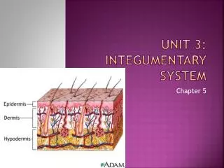
Unit 3: Integumentary System
Unit 3: Integumentary System. Chapter 5. What is integument?. Integument/Integument system: Debated as to whether or not your skin is an organ or organ system Accounts for 16% of body weight 2 Major Components: 1-Skin or Cutaneous Membrane Epidermis Dermis Hypodermis

Unit 3: Integumentary System
E N D
Presentation Transcript
Unit 3: Integumentary System Chapter 5
What is integument? • Integument/Integument system: Debated as to whether or not your skin is an organ or organ system • Accounts for 16% of body weight • 2 Major Components: • 1-Skin or Cutaneous Membrane • Epidermis • Dermis • Hypodermis • 2-Accessory Structures • Nails • Exocrine glands
Functions of Integumentary System • Protection • Excretion (salts, water, organic wastes) • Maintenance of body temp • Synthesis of vitamin D3 • Storage of nutrients (adipocytes) • Detection of touch, pressure, pain, etc.
Cutaneous membrane • Skin • Epidermis (superficial) • Dermis • Hypodermis (deep)
Epidermis • Thin skin=made of 4 layers of stratified Squamous tissue • Most of body • Thick skin=made of 5 layers • Hands and feet
Layers of Epidermis Made of Stratified Squamous epithelium • Stratum Corneum (superficial) • Stratum Lucidum • Stratum Granulosum • Stratum Spinosum • Stratum Basale or Germinativum (deep) • Cells eventually pass through all layers or can eventually be found
Stratum Corneum • Surface skin (most superficial) • Highly keratinized: thick, interwoven • 15-30 days to get from stratum basale to stratum corneum • Stay at stratum corneum for 2 weeks b/f shed • Loose 500 mL (1 pint) water a day through skin • Blisters: water retention between dermis/epidermis under high stress
Stratum Lucidum • Found in thick skin • Glassy, tough • Filled with keratin • Fibrous proteins—make up your hair and nails • Layer missing in “thin” areas
Stratum Granulosum • “Grainy Layer” • 3-5 layers • Have stopped dividing at this time • High in keratin • Makes cells flatter and thinner
Stratum Spinosum • “Spiny Layer” • 8-10 layers • Langerhans Cells =participate in immune response
Stratum Basale • Innermost • Closest to basement membrane • Sends projections into dermis (below basement membrane) • Called epidermal ridges • Give skin the whorls of fingertips • Basal cells =stem cells to make more skin • Merkel Cells =sense touch where no hair is present • Melanocytes =skin tone
Skin Color • Due to interaction of Epidermal Pigmentation and Dermal Circulation
Epidermal Pigmentation • 2 Pigments that control your skin color • 1-Carotene (orange-yellow) • Found in stratum corneum • 2-Melanin (brown, yellow-brown, black) • Found in stratum basale • protects us from UV radiation • Melanocytes produce • Dark skin = increased amount of Melanin production not increased amount of individual Melanocytes
Dermal Circulation • Gives pale or flushed look • Better circulation =flushed • Reduced circulation=pale
Why UV-Radiation is bad! • Damages DNA of the cell, causing mutations and promoting cancer development • Read bottom of 147(Melanocytes-Dermal Circulation)
Why UV-Radiation is good! Vitamin D3 • When exposed to sun, epidermal cells make D3, then the liver converts D3, and the kidney makes calcitriol. • Calcitriol=ability to absorb calcium and phosphorous (no calcitriol=impaired bone maintenance and growth)
Epidermal Conditions • Freckles • Areas where melanocytes are producing a higher than normal rate of melanin. • Birthmarks • Non-vascular • Overgrowth of melanocytes • Tattoo’s
The Dermis • Papillary Layer • Areolar tissue, capillaries, sensory neurons • Supplies epidermis 2. Reticular Layer • Collagen and elastic fiber
Wrinkles and Stretch Marks • Collagen fibers=strong, resists stretching BUT bend easily • Elastic fibers=stretch and return =flexible, elastic dermis • Aging, hormones, and excess UV = weakened fibers Wrinkles • Excessive stretching past fibers capabilities lead to damaged fibers=stretch marks • Caused by: pregnancy, major weight loss/gain • SOLUTION: Retin-A from vitamin A increases blood flow to dermis which increases chances for repair
Accessory Structures-Hair • 5 million hairs • 98% not on head • Hair Follicle=organ when hair is grown • Hair Root=anchors hair to skin • Hair Shaft=part you see
Accessory Structures-Hair • Is hair living? • No-Comprised of non-living cells • Hair follicles are • How is color determined? • Pigment produced by melanocytes • The biochemistry of these structures is affected by DNA • Hormonal/Environmental affects
Functions of Hair • Protection (eye lashes, head, sensory, ears, nose) • Root hair plexus=sensory nerve around each hair follicle • Arrector Pilli-smooth muscle attached to hair follicle=when stimulated, contracts, causes “goose bumps” • Stimulated by emotional states, response to cold
Growth and Replacement of Hair • Hair growth cycle=2-5 years • .33mm/day • Hair loss occurs when the follicle becomes inactive and shrinks • Over time, the connection breaks=hair loss • The old hole sheds, new one forms
Hair: Real Life Application • Male Pattern Baldness • Decrease in hormones circulating in the blood • Alopecia • 1 in every 100,000 • Complete hair loss all over body • Genetic • Causes death of hair follicles
Accessory Structure-Glands in the Skin • Sebaceous (Oil Glands-Holocrine) • Share a duct with hair • Waxy, oil secretions • Apocrine Sweat Glands (armpits, around the nipples, groin) • Odorous, sticky • Through hair follicle • Begin at puberty • MerocrineSweat Glands (all other sweat) • 2-5 million • High numbers in palms/soles
Accessory Structure-Nails • Provides protection to finger • Nail Body cover nail bed • Production at nail root • Lunala is pale=lack of blood vessels • Is dead, tightly compressed keratin packed cells
The Hypodermis or subcutaneous • Not part of integument but important for stabilization • Areolar/Adipose tissue • Elastic • Area for injections