ANTIGEN ANTIBODY REACTION
110 likes | 340 Vues
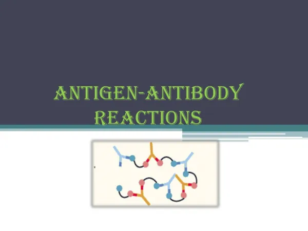
ANTIGEN ANTIBODY REACTION. By. Dr. Emad AbdElhameed Morad. Lecturer of Medical Microbiology and Immunology. Precipitation. It is a form of antigen antibody reactions in which the antigen is soluble. Types: this reaction can be done in:. Tube. Agar gel. Precipitation in tube.

ANTIGEN ANTIBODY REACTION
E N D
Presentation Transcript
ANTIGEN ANTIBODY REACTION By Dr. Emad AbdElhameed Morad Lecturer of Medical Microbiology and Immunology
Precipitation • It is a form of antigen antibody reactions in which the antigen is soluble. • Types: this reaction can be done in: Tube Agar gel
Precipitation in tube • When solutions of the antigen and antibody are mixed in tubes, cloudiness or turbidity occur.
Precipitation in agar • Example: Elek’s test. • A strip of filter paper saturated with diphtheria antitoxin is embedded in the agar plate. • Diphtheria is inoculated at right angles to the filter paper, then plates are incubated for two days at 37 degree. • If the organism is toxigenic, fine white lines of precipitation occur commencing from the streak.
Toxin antitoxin neutralization • Example: demonstrating toxigenicity of diphtheria. • Injecting two guinea pigs with the diphtheria isolate. • If the guinea pig protected with the antitoxin survives, while the unprotected one dies, the isolate is considered toxigenic.
Complement fixation test • The serum of the patient (antibody) is added to a known antigen and then complement is added. • If the serum contains antibodies to this antigen, they unite together and fix the complement. • If the serum has no antibodies, the complement is left free in the solution. • The second step is addition of sheep red blood cells coated with their antibodies. • In negative sera, the complement is free so it will cause hemolysis of red cells. • In positive sera, the complement is fixed and no hemolysis occurs.