Pleura & Lungs
860 likes | 1.18k Vues
Pleura & Lungs . Dr. Sarwar Hossain Khan. The Pleura. Is a sac on either sides of the mediastinum. Are occupied by the lungs by invagination. Is a serous membrane, lined by the mesothelium. Has 2 layers—parietal & visceral layers.

Pleura & Lungs
E N D
Presentation Transcript
Pleura & Lungs Dr. Sarwar Hossain Khan
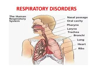
The Pleura Is a sac on either sides of the mediastinum. Are occupied by the lungs by invagination. Is a serous membrane, lined by the mesothelium. Has 2 layers—parietal & visceral layers. Is continuous with each other at the hilum of the lung. The 2 layers enclose between them the pleural cavity. This cavity is a site for—pneumothorax, pleural effusion,empyema & haemothorax.
Cont. • Pulmonary pleura—covers the surface of lung ,is free at the hilum & along the pulmonary ligament. • Cont. with the parietal pleura. • Parietal pleura—divided into costal,diaphragmatic,mediastinal & cervical. • Cervical—covers the apex of the lung & lies about 1 inch above the medial 1/3rd of the clavical. • Is covered by the sibson’s fascia.
PULMONARY LIGAMENT • Is a fold of the parietal pleura. • Around the hilum. • Acts as a dead space for the expansion of the veins. • Contains lymphatics & areolar tissues. • RECESSES: are folds of the parietal pleura making room for expansion of the lungs during breathing. • Costomediastinal & costodiaphragmatic.
CLINICAL IMPORTANCE • These get filled by pleural effusion when diseased. • SURFACE MARKING: • Cervical pleura • Anterior margin • Inferior margin • Posterior margin
Nerve supply • Parietal—intercostal & phrenic nerves • Is pain sensitive. • Pulmonary pleura—autonomic nerves—sympathetic nerves. • Not pain sensitive.
BLOD & LYMPHATIC SUPPLY • Parietal—intercostal,internal thoracic and musculophrenic arteries. • Veins drain into azygos and internal thoracic veins. • Lymphatics—intercostal, internal mammary & diaphragmatic nodes. • Pulmonary pleura—bronchial arteries & bronchopulmonary nodes.
APPLIED • Thoracocentesis • Pleurisy • Effusion • Pneumothorax • Haemothorax • Hydropneumothorax • empyema
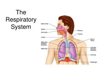
LUNGS • Organs of respiration • Mediastinum separates the 2 lungs • Spongy, grey, dark later on. • 600-700gm. R>L • Has apex, base, 3 borders, 2 surfaces. • Apex—above the level of the anterior end of the 1st rib—1 inch above the medial 1/3rd end of the clavicle. • Covered by sibson,s fascia.
Base—lies on the diaphragm. • Anterior border is thin—vertical on the R. • Ant. Border of L lung is notched—cardiac notch. • Here the heart is not covered by the lung & the pericardium. • Post.border is thick & corresponds to the heads of the ribs. • Extend from 7th C vert. to the 10th T vert.
Inferior border—separates the base from the costal & medial surfaces. • Costal surface. • Medial surface—post. & anterior parts. • Has numerous impressions.
Fissures & lobes. • R Lung—3 lobes—2 fissures; oblique & horizontal. • L Lung—2 lobes; 1 fissure. • Oblique fissure—cuts the whole thickness of the lung, except at the hilum. • Crosses post. Border, 2-3 inches below the apex. • Crosses inf. Border 2 in. from the medial plane.
Cont. • Horizontal fissure of R lobe divides the middle lobe from the upper lobe. • L lobe has a lingula below the cardiac notch. • No. of lobes may vary.
ROOTS OF THE LUNG • A pedicle connecting the medial surface of the lung with the mediastinum. • Short & broad. • Various structures exit & enter here from & to the lungs. • Lies opposite 5th-7th T vertebra.
contents • L—1-brochus, R-2-bronchi • 1 PA each. • 2 PV each. • Bronchial art.—2 on L, 1 on R • BV • Pulmonary plexuses—ant,post. • Lymphatics, LN, etc.
2 bronchi branch out from the trachea. • At the lower end of the 4th T vertebra. • R bronbus is short, wide,straight. • Infections are common.
Enters lungs through the hilum. • Divides into lobar bronchi—3 on R, 2 on L.
These further divide into—tertiary or segmental bronchi –10 on the R, 8 on the L(10) • These further divide into terminal bronchioles & these into respiratory bronchioles which aerates the pulmonary unit formed by—alveolar ducts,atria, air saccules, & pulmonary alveoli.
Arteries,veins, lymphatics & nerve supply. • Artery—bronchial art.—R side, one art.-either from 3rd post. Intercostal or upper L bronchial art. • L side-2 in no. from desc. Aorta. • Pulmonary art. –deoxygenated blood. • PV carry oxygenated blood to the heart.
veins • 2 bronchial veins on each side • R bronchial veins drain to azygos vein, • L veins-- into hemiazygos or the superior intercostal veins. • Drain blood from the 1st 1-2 divisions of the bronchi. • Rest of the blood is drained by the pulmonary veins.
lymphatics • Bronchopulmonary nodes.—from superficial & deep vessels • Some connection bet. the 2 groups exists. • NERVES • Parasympathetic—vagus—motor,sensory & secretomotor. • Sympathetics—T2-T5—bronchodilation. • Form plexuses around the hilum.
Bronchopulmonary segments • Sector of a lung that is aerated by a tertiary bronchus. • Pyramidal in shape with apex towards root. • There are 10 on the R, & 8 on the L. • Intersegmental planes—bet. The segments.