Advanced Diffraction Data Analysis with DIALS: A Modular Open Source Framework
DIALS (Diffraction Integration for Advanced Light Sources) is an open-source software toolbox designed for comprehensive diffraction data analysis. Developed collaboratively, DIALS provides a modular framework that facilitates the implementation of novel algorithms as well as the application of established methods to diffraction data. By incorporating features like sophisticated image handling, background correction, spot finding, and 3D integration, DIALS enhances the precision of crystallographic analysis for complex systems, supporting various detectors and refining parameters crucial for accurate data interpretation.

Advanced Diffraction Data Analysis with DIALS: A Modular Open Source Framework
E N D
Presentation Transcript
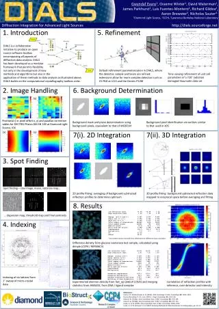
Gwyndaf Evans1, Graeme Winter1, David Waterman2, James Parkhurst1, Luis Fuentes-Montero1, Richard Gildea1, Aaron Brewster3, Nicholas Sauter3, 1Diamond Light Source, 2CCP4, 3Lawrence Berkeley National Laboratory Diffraction Integration for Advanced Light Sources http://dials.sourceforge.net 1. Introduction 5. Refinement DIALS is a collaborative initiative to produce an open source software toolbox encompassing all aspects of diffraction data analysis. DIALS has been developed as a modular framework that permits flexibility not only in the development of new methods and algorithms but also in the application of these methods to data analysis as illustrated above. DIALS builds on the computational crystallography toolbox cctbx. Default refinement parameterization in DIALS, where the detector, sample and beam are refined: extensions allow for more complex detectors such as CS-PAD at LCLS and the Dectris P12M Time varying refinement of unit cell parameters of a 720° radiation damaged thaumatin data set 2. Image Handling 6. Background Determination (x) (y) Positional (i.e. pixel offset x, y) and parallax correction tables for DECTRIS Pilatus 6M SN 100 at Diamond Light Source, I04 Background mask and plane determination using background pixels, equivalent to that of MOSFLM Background pixel identification via outliers similar to that used in XDS (x) (y) 7(i). 2D Integration 7(ii). 3D Integration 3. Spot Finding Spot finding – raw image, mean, variance map… 2D profile fitting: averaging of background subtracted reflection profiles to determine optimum 3D profile fitting: background subtracted reflection data mapped to reciprocal space before averaging and fitting 8. Results Low resolution limit 71.03 71.03 1.32 High resolution limit 1.30 7.12 1.30 Rmerge(all I+ and I-) 0.087 0.037 0.887 Rmeas(all I+ & I-) 0.096 0.040 0.982 Rpim(all I+ & I-) 0.040 0.017 0.415 Total number of observations 649690 4514 30623 Total number unique 115431 793 5655 Mean((I)/sd(I)) 10.6 34.4 1.7 Mn(I) half-set CC(1/2) 0.998 0.999 0.688 Completeness 100.0 100.0 99.9 Multiplicity 5.6 5.7 5.4 R* 23.50 Rfree* 24.31 *non protein atoms removed from refinement for difference map calculation … dispersion map, threshold map and final centroids 4. Indexing Difference density from glucose isomerase test sample, calculated using dimple (CCP4 / REFMAC5) Low resolution limit 42.40 42.40 1.32 High resolution limit 1.30 7.12 1.30 Rmerge(within I+/I-) 0.064 0.023 1.154 Rmeas (within I+/I-) 0.064 0.023 1.167 Rpim(within I+/I-) 0.009 0.004 0.172 Total number of observations 798525 5592 37737 Total number unique 9366 85 451 Mean((I)/sd(I)) 55.2 220.9 5.9 Mn(I) half-set CC(1/2) 1.000 1.000 0.962 Completeness 99.9 99.8 99.4 Multiplicity 85.3 65.8 83.7 Anomalous completeness 100.0 100.0 99.7 Anomalous multiplicity 46.6 56.1 44.1 DelAnom correlation half-sets 0.977 0.993 0.110 Mid-Slope of AnomProbability 2.102 Indexing of six lattices from 1° sweep of micro-crystal data Experimental electron density from fast_ep (SHELX C/D/E) and merging statistics from AIMLESS, from DNA / ligand complex Correlation of reflection profiles with reference, over detector and intensity References: Evans, P. R. & Murshudov, G. N. (2013). ActaCrystallogr. D. Biol. Crystallogr.69, 1204–1214. Grosse-Kunstleve, R. W., et al. (2002). J. Appl. Crystallogr.35, 126–136. Kabsch, W. (2010a). ActaCrystallogr. Sect. D Biol. Crystallogr.66, 133–144. Kabsch, W. (2010b). ActaCrystallogr. Sect. D Biol. Crystallogr.66, 125–132. Leslie, A. G. W. (1999). ActaCrystallogr. Sect. D Biol. Crystallogr. 1696–1702. Leslie, A. G. W. & Powell, H. R. (2007). Evolving Methods for Macromolecular Crystallography, pp. 41–51.