Impact of RNA Editing on Conformational Properties of 5HT2C Receptor's Intracellular Loop
This study investigates the conformational differences in the second intracellular loop (IL2) of 5HT2C receptors impacted by adenosine-to-inosine RNA editing. Analyzed through biased sampling Monte Carlo simulations, our findings reveal that edited receptor isoforms exhibit altered activity, with evidence of a reduced capacity for G-protein coupling. Specifically, differences in IL2 orientation between edited and unedited versions suggest a nuanced mechanism for receptor signaling in serotonin pathways, potentially influencing therapeutic strategies targeting these receptors.


Impact of RNA Editing on Conformational Properties of 5HT2C Receptor's Intracellular Loop
E N D
Presentation Transcript
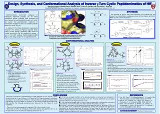
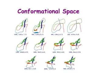
FIGURE 3: P1 and P2 corresponds to the alpha carbon of residues A3.53 and R4.39 M is the middle point in the vector P1P2 P3is C alpha of residue 4.35 is the vector corresponding to TM4 axes Vector MO is the projection of vector MP3 over the plane defined by vectors MP1 and . This vector is displayed just to emphasize the fact that vector MP3 is not in the same plane as MP1 and Positive values of Q are considered those which are at the left of R; negative values point to the right (Figure 3, from the viewer perspective). DIFFERENCES IN CONFORMATIONAL PROPERTIES OF THE SECOND INTRACELLULAR LOOP (IL2) IN 5HT2C RECEPTORS MODIFIED BY RNA EDITING CAN ACCOUNT FOR THE SILENCING OF CONSTITUTIVE ACTIVITY I. Visiers*, S. Hassan, H. Weinstein Dpt. Physiology and Biophysics. Mount Sinai School of Medicine, New York * e-mail address; irache@inka.mssm.edu ABSTRACT Adenosine-to-inosine RNA editing events demonstrated for 5HT2C receptors result in an alteration of the amino-acid sequence at positions 156, 158 and 160 of the receptor’s IL2. The “edited” receptor isoforms have reduced basal activity, but similar maximal responses to agonist binding. It has been proposed that the primary effect of editing may be to alter the ability of spontaneous isomerization to the active R* conformation rather than affecting the intrinsic ability of receptor isoforms to promote G-protein coupling. We have carried out conformational studies of the IL2 isoforms using Biased Sampling Monte Carlo simulations with a Screened Coulomb Potential-based Implicit Solvent Model, to compare the conformational space accessible to these loops in comparison to the unedited version. Our results show that the IL2 of the unedited (5HT2C-INI) receptor has a slightly larger population of structures oriented towards the 7TM bundle than the 5HT2C-VGV edited receptor. This difference in preferred orientation can affect the association of IL2 with other intracellular loop domains involved in G protein coupling, which implies a high sensitivity of the system to small changes in the interaction surface presented to other intracellular loops, and/or the G-protein. 2. The average angle value is slightly different for the populations of edited and unedited structures. However they are close indicating a significant extent of spatial overlap. <QINI>=-21.2 <QVGV>=-27.5 INTRODUCTION The 2C subtype of serotonin receptors (5HT2C) is a member of the G protein coupled receptor superfamily of seven transmembrane helix proteins. Residues in the second intracellular loop (IL2) connecting helices 3 and 4 in GPCRs has been demonstrated in numerous studies in GPCRs to play an important role in G-protein coupling [1-9]. IL2 and the third intracellular loop (IL3) have been shown to be required at the same time to achieve full G protein activaton, suggesting that they act in synergy [4], [10], [11]. Recently a number of naturally occurring adenosine-to-inosine RNA editing events were demonstrated for the 5HT2C receptor [12] [13]. They result in an alteration of the amino-acid sequence at positions 156, 158 and 160 in the second intracellular loop (IL2)(FIGURE 1). Pharmacological characterization found a decrease in agonist potency in the edited receptor 5HT2C-VGV in which the INI sequence was replaced by VGV. Competition binding experiments revealed that only non-edited receptors 5HT2C-INI had a serotonin high affinity state. Together these results indicate that the edited receptor couples less efficiently to G proteins. It has been suggested that this editing mechanism of 5HT2C receptors may represent a regulatory mechanism of receptor signal transduction at serotonergic synapses [14]. To identify the mechanistic basis for the altered properties of the edited 5HT2C receptor, we have investigated the effect of the mutations on the conformation of IL2 using a Biased Sampling Monte Carlo simulation. The resulting conformations were examined in a receptor model to identify the potential role of editing on the functional role of IL2. We find that the differences in the population of conformations between the edited and unedited forms relate to the orientation of the loop relative to the interior of the 7 transmembrane helix and IL3. Given the role of the association of IL2 with other intracellular loop domains in G-protein coupling, these differences in the population of IL2 conformations explain the effect of loop editing in the coupling efficiency of the edited receptors. The unedited receptor has an average Q in the range of -21.2 . This area is the most populated one in the case of the WT receptor with 41.7% of the structures, while the same area in the case of the edited receptor has a 31.1% population. However the most populated region in the edited receptor is the one corresponding to the third bin -25>Q>50 (41.6%). The area corresponding to Q>0 is also larger for the WT than for the edited receptor. The WT loop shows a clear trend toward higher values of Q than the edited one. This indicates that the non-edited (5HT2C-INI) receptor has a slightly larger population of structures oriented towards the 7TM bundle than the 5HT2C-VGV edited one. Those structures oriented towards the interior of the bundle would also have greater spatial proximity to the third intracellular loop (IL3). GEOMETRIC CHARACTERIZATION OF THE CONFORMATIONAL SPACE IL3 cartoon FIGURE 4: Same group of structures as in figure 2, viewed from the intracellular side of the receptor in relation to IL3 shown schematically. This figure shows the trend in the unedited receptor to locate IL2 closer to IL3. The values of Q for the 130 and 150 conformations of WT and VGV mutant respectively were sorted and plotted in graphs 1 and 2 along with the energy of each particular structure. EDITED RECEPTOR WILD TYPE CONCLUSIONS: 1. The non-edited (5HT2C-INI) receptor has a slightly larger population of structures oriented towards the 7TM bundle than the 5HT2C-VGV edited one. Those structures oriented towards the interior of the bundle would also have greater spatial proximity to the third intracellular loop. Our results indicate the likelihood of a direct interaction between IL2 and IL3, and thus provide a molecular basis for the observed synergistic effect of the two loops in receptor coupling to G proteins. 2. The conformational effect of the substitution of residues I156, I158 and N160 by VGV is a change in the distribution of structures covering the accessible conformational space. While the distribution of the edited loops exhibits some overlap with that of the unedited version, there is a clear deviation away from the region of potential interactions with IL3. This may explain the fact that the 5HT2C-INI non-edited receptor couples more efficiently to G proteins while retaining Vmax. 3. Our results support the hypothesis that a small difference in loop orientation is sufficient to account for the observed reduction in G-protein coupling by disrupting the optimal orientation of IL2 relative to IL3 that achieves binding with their respective sites at the G protein. This implies a high sensitivity of the system to small changes in the interaction surface presented to the IL3 and/or the G-protein. ANGLE ENERGY(Kcal) ENERGY(Kcal) ANGLE FIGURE 1: IL2 loop peptides used in this study. Positions 156, 158 and 160 where editing events occur are highlighted. Throughout the simulation the position of the fragments 3.53 to 3.55 and 4.38 to 4.39 (red filled circles) are harmonically constrained to fit the position of the cytoplasmic ends of TM3 and TM4 by constraining the phi and psi dihedral angles to remain helical, as well as by restraining interesidue distances between the two helical fragments to correspond to the receptor model. Graph 1 Graph 2 EXPLORATION OF THE CONFORMATIONAL SPACE The conformational space of the loop was explored with the Biased Sampling Monte Carlo method of Conformational Memories (CM) described earlier [15], [16]. Each exploration of the conformational space involves 130 runs of simulated annealing for WT and 150 for the edited receptor, yielding 130 and 150 initial structures for each isoform of the 5HT2CR. The aqueous environment of the loop peptide is modeled using an efficient Implicit Solvent Model (ISM) that is based on a Screened Coulomb Potential formulation (the SCP-based ISM) (poster 1491). The CHARMM forcefield was used for the energy calculations. Positive values of Q identify conformations where the C alpha of residue 4.35 is oriented closer to the interior of the bundle. The more negative the value of the angle, the more the loop “swings” away from the interior of the bundle. En is the total energy and runs from 1 to 130 for WT and 1 to 150 for the mutant K is Boltzman constant T is 300°K Z is the partition function. REFERENCES: 1.Ballesteros, J., S. Kitanovic, F. Guarnieri, P. Davies, B.J. Fromme, K. Konvicka, L. Chi, R.P. Millar, J.S. Davidson, H. Weinstein, et al., Functional Microdomains in G-protein-coupled Receptors: The conserved arginine cage motif in the gonadotropin-releasing hormone receptor. J. Biol. Chem., 1998. 273(17): p. 10445-53. 2.Franke, R.R., B. Konig, T.P. Sakmar, H.G. Khorana, and K.P. Hofmann, Rhodopsin mutants that bind but fail to activate transducin. Science, 1990. 250: p. 123-125. 3.Franke, R.R., T.P. Sakmar, R.M. Graham, and H.G. Khorana, Structure and function in rhodopsin. Studies of the interaction between the rhodopsin cytoplasmic domain and transducin. J Biol Chem, 1992. 267(21): p. 14767-74. 4.Konig, B., A. Arendt, J.H. McDowell, M. Kahlert, P.A. Hargrave, and K.P. Hofmann, Three cytoplasmic loops of rhodopsin interact with transducin. Proc Natl Acad Sci U S A, 1989. 86(18): p. 6878-82. 5.Farahbakhsh, Z.T., K. Hideg, and W.L. Hubbell, Photoactivated conformational changes in rhodopsin: a time-resolved spin label study. Science, 1993. 262(5138): p. 1416-9. 6.Farahbakhsh, Z.T., K.D. Ridge, H.G. Khorana, and W.L. Hubbell, Mapping light-dependent structural changes in the cytoplasmic loop connecting helices C and D in rhodopsin: a site-directed spin labeling study. Biochemistry, 1995. 34(27): p. 8812-9. 7.Kuhn, H. and P.A. Hargrave, Light-induced binding of guanosinetriphosphatase to bovine photoreceptor membranes: effect of limited proteolysis of the membranes. Biochemistry, 1981. 20(9): p. 2410-7. 8.Moro, O., J. Lameh, P. Hogger, and W. Sadee, Hydrophobic amino acid in the i2 loop plays a key role in receptor-G protein coupling. J Biol Chem, 1993. 268(30): p. 22273-6. 9.Arora, K.K., A. Sakai, and K.J. Catt, Effects of second intracellular loop mutations on signal transduction and internalization of the gonadotropin-releasing hormone receptor. J Biol Chem, 1995. 270(39): p. 22820-6. 10.Cypess, A.M., C.G. Unson, C.R. Wu, and T.P. Sakmar, Two cytoplasmic loops of the glucagon receptor are required to elevate cAMP or intracellular calcium. J Biol Chem, 1999. 274(27): p. 19455-64. 11.Wong, S.K., C. Slaughter, A.E. Ruoho, and E.M. Ross, The catecholamine binding site of the beta-adrenergic receptor is formed by juxtaposed membrane-spanning domains. J Biol Chem, 1988. 263(17): p. 7925-8. 12.Herrick-Davis, K., E. Grinde, and C.M. Niswender, Serotonin 5-HT2C receptor RNA editing alters receptor basal activity: implications for serotonergic signal transduction. J Neurochem, 1999. 73(4): p. 1711-7. 13.Niswender, C.M., S.C. Copeland, K. Herrick-Davis, R.B. Emeson, and E. Sanders-Bush, RNA editing of the human serotonin 5-hydroxytryptamine 2C receptor silences constitutive activity. J Biol Chem, 1999. 274(14): p. 9472-8. 14.Burns, C.M., H. Chu, S.M. Rueter, L.K. Hutchinson, H. Canton, E. Sanders-Bush, and R.B. Emeson, Regulation of serotonin-2C receptor G-protein coupling by RNA editing [see comments]. Nature, 1997. 387(6630): p. 303-8. 15.Guarnieri, F. and S.R. Wilson, Conformational Memories and a Simulated Annealing Program That Learns: Application to LTB4. J. Comput. Chem., 1995. 16(5): p. 648-653. 16.Guarnieri, F. and H. Weinstein, Conformational memories and the exploration of biologically relevant peptide conformations: An illustration for the gonadotropin-releasing hormone. J. Amer. Chem. Soc., 1996. 118: p. 5580-5589. FINDINGS 1. The exploration of the conformational space converged for both the WT and the edited loop. The population of different regions of the space is calculated by parsing the conformational space described by Q into 4 bins of 25 degrees and calculating the Boltzman probability for each one of the bins. The first 90 runs provided 90 structures, and the results of successive runs are added in until convergence. Convergence is achieved when the population of each bin changes less than 1% upon addition of structures from a subsequent run. FIGURE 2: The resulting structures were oriented by superposition of the backbone of residues 3.53 to 3.55 and 4.38 to 4.39 in the 5HT2CR model and grouped in clusters with a backbone RMS <1.57. Representative structures were obtained for each cluster. These are shown in the figure. The red arrow indicates the area covered by the “swing” of the loop. WILD TYPE EDITED