Download
1 / 32
320 likes | 354 Vues
Discover the world of microorganisms, from microscopic life forms to infectious agents, in a multidimensional exploration of biology. Learn about the major groups of microbes, their classification, and the vital role they play in ecosystems and human civilization.

E N D
MICRB 106/107Elementary Microbiology & Lab Read both Contracts! Finding Me Goals & Objectives Evaluation Criteria Policies, Requirements, etc… Textbooks The Schedules
MICRB 106: Evaluation • Participation (9%) • Self-evaluation • Professor’s evaluation • Quizzes (4 x 2% ea. = 8%) • Unannounced. • Only on Mondays. • Exams I (15%) • Exams II, III, IV (20% ea. = 60) • Bad Bug Talk (8%) • With a partner. • 15 minute maximum.
MICRB 107 (lab): Evaluation • Lab Assignment Worksheets (50%) • Five 10% submissions. • Due dates announced. • Quizzes (6 x 2% ea. = 12%) • Unannounced. • Only on Wednesdays. • Practical Exams (2 x 15% ea. = 30%) • Lab Behavior/Etiquette (8%)
“Microbiology is a branch of Biology that uses procedures involving sterile technique and the use of culture media which are necessary to isolate and grow microorganisms” – Roger Stanier, 1978 • “Microorganisms (microbes) are organisms too small to be seen clearly by the naked eye” - Prescott et al., 2001
Which are the microorganisms? • Life forms, or other self replicating entity, that requires microscopy technology to be clearly visualized. • Many are unicellular, sometimes cells are organized in filaments or clumps, and others are complex with only a portion of their life cycle being microscopic. • Most can carry out life processes independently from other cells, others are highly parasitic. • They often require specialized techniques for their study: microscopy, culturing, biochemical and/or molecular. • They include all prokaryotic and many eukaryotic life forms.
We’ll get back to these differences in more detail in a latter lecture. What are some of the major groups of life that we call microbes? Large difference in scale. Average eukaryotic cell is about 50x bigger than the average prokaryote.
3 Domains of Life (2 Prokaryote & 1 Eukaryote) “Tree of Life” Most all of life’s diversity is microbial; represented in all major phyla.
Don’t forget, some animals too! Flatworms & Roundworms Trichinella spiralis larva in skeletal muscle (W.M., X260). The spiral juvenile and its nurse cell are visible in this preparation.
Viruses: An infectious particle with an acellular organization of protein and nucleic acids (RNA or DNA), and lacking independent metabolism. It requires the metabolism of a host cell in order to replicate. Viruses are about 50 to 200 nm in size. Prion: An infectious aberrant brain protein that causes abnormal aggregation of similar normal brain proteins; no nucleic acids.
Note on Classification: Prokaryote (e.g. E. coli) • Domain: Bacteria • (no kingdom) • Phylum: Proteobacteria • Class: γ-proteobacteria • Order: Enterobacteriales • Family: Enterobacteriaceae • Genus: Escherichia • Species: coli Eukaryote (e.g. Humans) • Domain: Eukarya • Kingdom: Animalia • Phylum: Chordata • Class: Mammalia • Order: Primata • Family: Hominidae • Group: Homo • Species: sapiens Binomial nomenclature: Genus species (italic or underlined) Just like varieties of apples, or races of people, there are strains of a prokaryote species (e.g. the harmless Escherichia coli K12 versus the deadly pathogenic E. coli O157:H7). Why so?
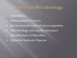
Why Study Microbes? 1) Microbes and Man in Sickness and Health • Parasitism; Pathogens (disease causing) • Infectious disease is leading cause of death in developing countries (45%). • Commensalisms; Natural Microbiota (do no harm) • Mutualisms; Natural Microbiota (do us good; probiotics) 2) Major Developments in Biology: • Recombinant DNA technology; Cloning • Industrial Applications (antibiotics) • Polymerase Chain Reaction (PCR) for Diagnostics • Genomics (Computer Technology for Studying Life) 3) The Role of Microbes in Ecosystems • Sources for drug discovery (antibiotics & antiviral drugs) • Cycling of Elements (Biogeochemistry of C, N, S …) • Agriculture (crop diseases; nutrient enhancement) • Pollution Bioremediation (oil, xenobiotics)
4) Microbes in Human Civilization: • Food and Beverage preservation (pre-history) • Turns in History - The “Four Horseman”: War, Famine, Pestilence, Death. • Moses leads Israelites out of Egypt; facilitated by plague (1500 BC) • Athenians (Greeks) lost Peloponnesian War in 404 BC due to plague (Yersinia pestis; bacterial disease) • Fall of Rome (565 AD) due to overcrowding exacerbated by Attila the Hun’s (barbarians’) army cutting off water supplies to Rome – epidemic malaria (protist disease) and other infectious diseases. • Spanish conquering the Aztec civilization (1500’s) by introducing small pox and measles (viral disease). • Salem Witch Hunt (1692): Puritan rye gets infected by Ergot (Claviceps purpurea; fungal plant pathogen); bread makes them loony; hallucinations perceived as evil spells. • Great Famine (Ireland ca 1850’s): potato blight (Phytophthora infestans; oomycete fungi) starved ten’s of thousands to death; over a million immigrated to America.
Over 3.5 billion years of “microbes”;only 400 years of Microbiology! • First life at 3.8 Ga by evolved chemical complexity; alien entry; or divine intervention. • Earth then was extreme: no oxygen, intense radiation, very hot, & electrical discharge. • Energy for earliest life from reduced (electron rich) organic and inorganic compounds. • Later came the ability to capture light energy for synthesis of organic matter from CO2. • Oxygenic photosynthesis slowly oxygenated the atmosphere; greater energy yield from metabolizing organic matter by aerobic respiration helped accelerate evolution.
Over 400 years of Microbe Hunting!What fundamental discoveries started it all? • First Light Microscopes (1600s) • Origins of New Life and the Debunking “Spontaneous Generation” (1660’s to 1860s) Louis Pasteur • Linking Microbes to Human Disease (late 1800s) Robert Koch • Discovering Vaccines and Immunity (late 1800s) Edward Jenner, Elie Metchnikoff , Emil von Behring • Agricultural and Environmental Microbiology (late 1800s) Martinus Beijerinck, Sergei Winogradsky
First Light Microscopes Zacharias Jannsen (1595), Robert Hooke (1665): first “cells” from cork; Micrographia, 1665
First Microscopes Antonie van Leeuwenhoek (1676): excellent simple microscopes; discovered and observed microbes, called them “animalcules”.
Size? See Table 3.1
Microscopy: • Light Microscopy • - Specimen illuminated with visible or ultraviolet light. • - Used for seeing specimens from 0.5 µm to 0.5 cm. (10,000-fold difference) • Electron Microscopy • Specimen “illuminated” with electrons. • Used for specimens from 0.5 nm to 50 µm. (100,000 – fold difference)
Light Microscopes: • Anatomy & Optics: • Light source is from underneath. • Condenser concentrates the light; forming a “cone shaped” beam pointing into the specimen. • Note that the image gets flipped after the sample. • The prism redirects the path of light to the oculars, during the path from prism to oculars the image flips again – back to “normal”. • Magnification: • Objective lenses • Ocular lenses • Total magnification is their product.
Blurring and dimming of the image at higher objective magnification (e.g. 100 x): • Light changes its pathway angle, or bends, when is passes through materials of different consistencies (optical density). This is called refraction. • It happens when light passes through glass into air. Thereby, some light illuminating a sample may not pass into the objective (hence dimming), or the path may be shifted out of focus (hence blurring). • The problem appear worse with a 100x objective. • Immersion oil between specimen and objective is the solution; it’s similar to glass.
Brightfield Illumination: * Uses full spectrum, or white light (“all the colors of the rainbow”), directed to pass through the specimen. * Often very small non-pigmented specimens makes them appear nearly featureless, transparent, even invisible. * Colored stains are used to enhance the image of very small specimens
Fluorescence Microscopy: • Another way to see cells and structures difficult to see with brightfield illumination, and more sensitive than colored dyes. • The microscope illuminates the sample with ultraviolet light (UV), and the sample must be dyed with a compound that will fluoresce a color when illuminated with UV light; call fluorochromes. • Immuno-fluorescence: • Antibodies are immune system proteins that can bind to a target specimen very specifically. They are very useful in fluorescent microscopy when they have a fluorescent dye bound to them.
Darkfield Microscopy: Brightfield Darkfield • The image is like a photographic negative of the brightfield image. • Stains are typically not used with darkfield. • Special optics enhance the contract so more details are seem. • Instead of a solid “cone of light” condensed onto the specimen, brightfield optics create a hollow cone that illuminate the specimen’s sides. Salamander egg; 500 µm Human cheek cells; 50 µm (stained)
Darkfield Optics The annular stop blocks light from most of the condenser lens, except for the edges; hence a “hollow cone” of light points onto the sample. Without the extra light coming through the sample, the background is dark.
Other Kinds of Light Microscopy • Phase Contrast:partly like darkfield, but the optics create a light wavelength shift as it passes through denser parts of the specimen; resulting in even greater contrast enhancement so to see even more detail. • Differential Interference Contrast (DIC):Uses two light sources condensing on the sample to create more contrast than phase contrast. Furthermore, the light passes through a prism, resulting in a separation of color and more color seen in the image. • Confocal Microscopy:Uses lasers and fluorochrome dyes. Laser illumination permits focusing on specific layers within a specimen. When coupled with computer software, 3D images can be generated.
Image Resolution: Light’s Limitation • Resolution refers to the ability to distinguish between two points in space. • Higher resolution means you distinguish between two objects that are closed together. • Maximum resolution is the closest two point in space can be apart from each and still distinguish them. • Illuminating a sample with a smaller wavelength of light can improve maximum resolution. UV light is a little better that white light, and electrons are the best!
White Light has a maximum resolution of about 0.2 µm. Two cells or viral particle smaller than this will appear as a single blurred object when next to each other.
Electron Microscopy The Maximum Resolution is 0.5 nm, which is 400-times better than light. Transmission EM creates 2D images, and specimens must be very thin for electrons to pass through. Scanning EM makes 3D images of any surface.
TEM (2D) SEM(3D)Both require special staining of a non-living specimen.
Be able to compare the differences between light and electron microscopy.