Incidental Findings in CT Angiography in Acute Stroke Patients and Treatment Changes: Single Center Observation
This retrospective study evaluated the percentage of incidental findings in CT angiography (CTA) scans performed on acute stroke patients. The study also assessed changes in patient management based on these findings. Results showed a high number of incidental findings, with 14% of patients having a change in treatment strategy.

Incidental Findings in CT Angiography in Acute Stroke Patients and Treatment Changes: Single Center Observation
E N D
Presentation Transcript
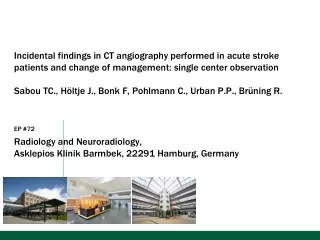
Incidental findings in CT angiography performed in acute stroke patients and change of management: single center observation Sabou TC., Höltje J., Bonk F, Pohlmann C., Urban P.P., Brüning R. EP #72 Radiology and Neuroradiology, AsklepiosKlinikBarmbek, 22291 Hamburg, Germany
Purpose In the diagnostic process of acute stroke in most centers computed tomography (CT) is established as a combination of unenhanced cranial CT, plus CT angiography and CT- based perfusion measurements. Aim of this retrospective evaluation was to evaluate the percentage of incidental findings during this CTA scans (from aortic arch), and to evaluate changes in therapy based on these observations.
Materials and Methods We have retrospectively selected from the PACS-Database 100 patients which underwent an unenhanced cranial CT examination and a CT-Angiography between 20.3. and 06.08.2012 (median age 74y, 31y-95y; 52 males, 48 females). CTA scan range was from the aortic arch to scull in all cases at an injection of 80 - 130 cc contrast (body weight adapted). The raw data (Philips Brilliance 40 and GE Optima 660) were reconstructed in axial, sagittal and coronal planes with a 2-3 mm slice thickness. Image acquisition was followed by a consensual evaluation and classification (resident and neuroradiologist) according to a predefined score (e.g. in category 1 for pulmonary lesions)= dissection, bronchial carcinoma, pulmonary embolism). From the hospital’s SAP database a possible change of patient management was retrospectively evaluated.
Results • The following secondary findings were observed in a total of 73/100 patients: • pulmonary section: 20% (for example pulmonary carcinoma, pulmonary artery embolism); • Vascular: 44% (i.e. dissection, figure 1), • intracranial findings: 19% (for example hypophyseal adenoma) • musculoskeletal findings: 11% (for example epidural hemaotma in the cervical spine, figure 3): • ENT-section: 5%. • Secondary findings which altered the treatment strategy such as central pulmonary embolism, acute pulmonary edema (figure 2a) or lung carcinoma (figure 2c) were observed in 14 patients.
Figure 1 86 years old female patient: the CT angiography shows a Stanford B aortic dissection with a partially thrombosed lumen (1a- 1c).
Figure 2 In the partially depicted, apical lung segments one could see secondary findings such as pulmonary edema (2a), pleural effusion with pneumonic infiltration (2b) and a lesion suspected to be tumor (2c),
Figure 3 In an 68 year old patient the CT scan revealed a space occupying lesion in the cervical spinal canal (3a, arrow); with suspicion of epidural hematoma in the MRI study next morning (3b)
Literature William B. Hall, MD; Sherstin G. Truitt, MD; Leslie P. Scheunemann, MD; Sidharth A. Shah, MD; M. Patricia Rivera, MD; Leonard A. Parker, MD; Shannon S. Carson, MD; The Prevalence of Clinically Relevant Incidental Findings on Chest Computed Tomographic Angiograms Ordered to Diagnose Pulmonary Embolism. Arch Intern Med. 2009;169(21):1961-1965. doi:10.1001/archinternmed.2009.360; Hoffstetter P1, Herold T, Daneschnejad M, Zorger N, Jung EM, Feuerbach S, Schreyer AG. Non-trauma-associated additional findings in whole-body CT examinations in patients with multiple trauma. Rofo. 2008 Feb;180(2):120-6. Epub 2007 Nov 16. Broderick JP, Palesch YY, Demchuk AM, et.al.; von Kalle T, Fabig-Moritz C, Heumann H, Winkler P. Incidental findings in paranasal sinuses and mastoid cells: a cross-sectional magnetic resonance imaging (MRI) study in a pediatric radiology department. Rofo. 2012 Jul;184(7):629-34. doi: 10.1055/s-0032-1312861. Epub 2012 May 22. Weber C, Grzyska U, Lehner E, Adam G. Clinical relevance of cranial CT under emergency conditions--basic neuroradiologic investigations. Rofo. 2003 May;175(5):654-62.
Conclusion A high number of examination in acute stroke patients yielded incidental findings within the examined region. In our single center analysis, in 14% patients had an alternative treatment strategy based on CT findings, especially chest findings. For questions please contact t.sabou@asklepios.com