ACUTE PANCREATITIS
1.02k likes | 1.17k Vues
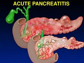
Acute pancreatitis is a non-bacterial inflammation of the pancreas, characterized by the activation and digestion of the gland by its own enzymes. It can present as mild parenchymal edema to severe hemorrhagic pancreatitis with necrosis. Common causes include gallstones, alcohol consumption, and hyperlipidemia, among others. Clinically, patients often experience abdominal pain, varying from mild discomfort to severe, toxic conditions. Understanding the etiology, pathogenesis, and clinical features is essential for effective management and treatment of this condition.

ACUTE PANCREATITIS
E N D
Presentation Transcript
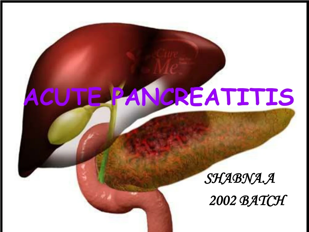
ACUTE PANCREATITIS SHABNA.A 2002 BATCH
DEFINITION • NON-BACTERIAL INFLAMMATION OF PANCREAS CAUSED BY ACTIVATION,INTERSTITIAL LIBERATION AND DIGESTION OF GLAND BY ITS OWN ENZYMES • MILD PARENCHYMAL OEDEMA→SEVERE HAEMORRHAGIC PANCREATITIS→GANGRENE AND NECROSIS
1) GALL-STONE PANCREATITIS • 40% of all cases • 90% of a/c pancreatitis • Transient obstruction of pancreatic duct by a gall stone in CBD at Ampulla of Vater • Brief obstruction • Middle→late forties • Women
2)ALCOHOLIC PANCREATITIS • 40% of all cases of pancreatitis • 75% of c/c pancreatitis • Diagnosed after patients have consumed alcohol for atleast 20 yrs(b/w 6-10 yrs),daily consumption averages 100-150 gm/day • Diet richer in fat & protien
MECHANISM OF ALCOHOL-RELATED INJURY • Alcohol is a known stimulant of gastric acid secretion→ duodenal acidification→ stimulates secretin release→ ↑exocrine secretion of pancreatic water & HCO3→parenchymal injury • Ethanol induces spasm of sphincter of Oddi → obstruction to pancreatic juice outflow
Ethanol→↑ed conc: of enzyme protein in pancreatic juice→ precipitation in pancreatic ducts→ multiple ductal obstruction • ↑es pancreatic duct permeability→ leaking of enzymes→ damage to pancreas • a/c ethanol ingestion→ ↓es pancreatic blood flow→ ischaemia→ cellular damage
3) HYPERCALCEMIC STATES • Commonly hyperparathyroidism • Favour intraductal precipitation of calcium stones • ↑es permeability of ducts→ enzyme leakage 4) HYPERLIPIDEMIA • With ↑ed chylomicrons & VLDL • Release of large amounts of toxic fatty acids by pancreatic lipase
5)HEREDITARY PANCREATITIS • Dominant trait • Mutation in gene for trypsinogen→ interferes with normal trypsin degradation→ digest pancreas • Rare • b/w 12-14 yrs • Recurrent a/c attacks
6)POST-OPERATIVE (IATROGENIC) PANCREATITIS • Occurs after so many procedures • Direct injury to the gland Pancreatic biopsy,Pancreatic resection • Obstruction of pancreatic duct Long-arm T-tube through sphincter,forceful dilatation of sphincter to >5 mm diameter
Follows endoscopic sphincterotomy • Follows surgical sphincteroplasty • Follows Billroth II gastrectomy
Definite cause Azathioprine Estrogen Probable cause Thiazide diuretics Furosemide Ethacrynic acid Sulfonamides Tetracycline L-asparginase Corticosteroids Phenformin Procinamide Valproate Clonidine Pentamidine 7)DRUGS
8) PANCREATIC DIVISUM • Failure of dorsal & ventral duct system to join • Young adulthood • Recurrent episodes of acute pancreatitis DORSAL PANCREAS VENTRAL PANCREAS
9) MISCELLANEOUS CAUSES • Follows scorpion venom poisoning • Infections(mumps, group B coxsackie viruses, herpes simplex, IMN) • Exposure to anticholinesterase insecticides 10) IDIOPATHIC • 15-20 % of total • Other causes should be ruled out before confirmation
1) OBSTRUCTION-SECRETION • Partial duct obstruction+Stimulation of pancreatic secretionMore severe pancreatic inflammation • Biliary pancreatitis gall stone obstructs pancreatic duct • A/c alcoholic pancreatitis Alcohol stimulates gastric acid secretion which then releases secretin stimulates pancreatic secretion • Alcoholspasm of sphincterobstructs pancreatic flow
2)COMMON-CHANNEL THEORY • Gall-stone in Ampulla of Vater obstructs CBD & pancreatic duct common channel b/w them bile reflux into pancreatic duct. • Infected bile contains deconjugated bile salts & bile incubated with pancreatic juice contains lysolecithine injure lining of pancreatic ducts es permeability leakage of pancreatic enzymes to surrounding parenchyma pancreatitis
3)DUODENAL REFLUX After Billroth II gastrectomy duodenal reflux occurs through papilla of Vaterpancreatitis 4)ED PANCREATIC DUCT PERMEABILITY Acute alcohol ingestion,direct exposure of duct to deconjugated bile salts,pancreatic secretion against an obstruction,acute hypercalcemia duct permeability enzyme leakage pancreatitis
Hemorrhagic Pancreatitis and Fat Necrosis Saponification chelation of Ca with fatty acids liberated by pancreatic enzymes
Changes due to action of pancreatic enzymes • Microvascular leakageoedema • Fat necrosis by lipase • Proteolytic destruction of pancreas • Destruction of blood vessels - interstitial hemorrhage
Milder– a/c interstitial pancreatitis or acute edematous pancreatitis • Histology • Interstitial edema • Focal areas of fat necrosis in pancreas & peripancreatic fat • No pancreatic necrosis
Severe –a/c necrotizing pancreatitis • Necrosis of pancreatic tissue affect acinar, ductal tissue & islets • Damage to vessel hemorrhage into parenchyma
Diverse spectrum of illness • Mild,short-lived,self-limiting illness severe toxic condition
ABDOMINAL PAIN • Predominant feature • Begins in mid-epigastrium • Penetrating,radiating to back • Also in right or left upper quadrant • Also as generalized non-localized pain • Variable intensity-less severe with oedematous than necrotizing form of disease
Alcohol associated pancreatitispain onset b/w 12-48 hours after an episode of inebriation Gall-stone associated pancreatitispain follows ingestion of a large meal nausea,vomiting & retching severe,protracted vomiting
RARE PRESENTATION • Severe systemic illness hypotension,hypoperfusion & mental depression {signs of profound fluid loss}
SIGNS • Fever(100-101ºF) • Tachycardia • Epigastric tenderness • Abdominal distension paralytic ileus from retroperitoneal phlegmon distension with intraperitoneal fluid in necrotizing pancreatitis
Guarding & rigidity • Bowel sounds ed or absent • No palpable mass usually • If palpable A swollen pancreas (phlegmon, pseudocyst or abscess)
Severe haemorrhagic pancreatitis In < 3% of patients • GREY-TURNER’S SIGN-bluish discolouration in left flank
CULLEN’S SIGN-bluish discolouration in periumbilical region • Results of tracking of blood-stained retroperitoneal fluid through tissue planes of abdominal wall to the flank or along the falciform ligament to the umbilical area • Indicates presence of severe episode of a/c haemorrhagic pancreatitis
Grey turner’s sign Cullen’s sign
Jaundice uncommon finding seen in patients with gall-stone pancreatitis also follow compression of distal CBD by oedema of head of pancreas • Major circulatory derangements in severe pancreatitis hypotension,hypoperfusion related to hypovolemia & ed preload to heart
Extraabdominal manifestations • Left pleural effusion or left hemidiaphragm elevation(1/3 patients) • Signs of a/c pulmonary failure tachypnoea,dyspnoea,cyanosis • Subcutaneous fat necrosis • Cerebral abnormalities confusion,psychosis & coma follow hyperosmolarity,hypoperfusion & hypoxia,cerebral fat embolism or DIC
DIFFERENTIAL DIAGNOSIS • Perforated peptic ulcer • Acute cholecystitis • Gangrenous small bowel obstruction • Cholangitis • Gastroenteritis • Vascular occlusion • Pancreatic cancer • Renal colic
SERUM AMYLASE • 90% of patients • Most widely used • Hyperamylasemia observed from 24 hrs to 7 days • es to > 2.5 times • Values > 1000 IU/dl Biliary pancreatitis • Lower values a/c alcoholic pancreatitis • P-type isoamylase more diagnostic • 10% have normal serum amylase level
2) SERUM LIPASE • More reliable to diagnose • Pancreas is the main source of lipase in blood • Duration of hyperlipasemia exceeds that of hyperamylasemia • Time consuming & difficult not preferred 3) SERUM TRYPSIN & ELASTASE CONCENTRATION • Sensitive markers of pancreatic inflammation • Radioimmunoassay kits are available
4) ADDITIONAL LAB TESTS • Haematocrit ed due to dehydration or ed due to pancreatic or retroperitoneal blood loss in necrotizing pancreatitis • ed WBC count >10000 cells/mm³ • Hyperglycemia,mild azotemia,hypocalcemia • LFT Normal or mild in S.bilirubin (< 2 mg%) Common in gall-stone pancreatitis Also due to partial obstruction CBD by swollen pancreatic head
5) RADIOLOGICAL PROCEDURES Supportive to clinical and lab diagnosis
Plain chest X-ray • Supportive of diagnosis • Left basal atelectasis, elevation of left hemidiaphragm, left pleural effusion • Reflect significant inflammatory process • Helpful to eliminate other diagnosis