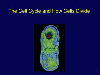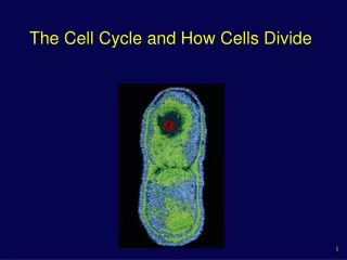
How Cells Divide Mitosis is a key phase of the cell cycle Phases of the Cell Cycle
How Cells Divide Mitosis is a key phase of the cell cycle Phases of the Cell Cycle. G1: is the primary growth phase of the cell. For many organisms, this encompasses the major portion of the cell’s life span. S: is the phase in which the cell synthesizes a replica of the genome.

How Cells Divide Mitosis is a key phase of the cell cycle Phases of the Cell Cycle
E N D
Presentation Transcript
How Cells Divide Mitosis is a key phase of the cell cycle Phases of the Cell Cycle
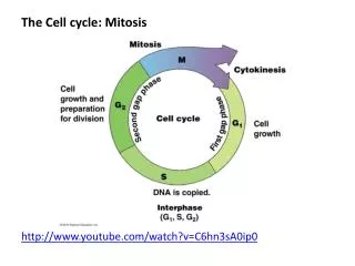
G1:is the primary growth phase of the cell. For many organisms, this encompasses the major portion of the cell’s life span. • S:is the phase in which the cell synthesizes a replica of the genome. • G2:is the second growth phase, in which preparations are made for genomic separation. During this phase, mitochondria and other organelles replicate, chromosomes condense, and microtubules begin to assemble at a spindle. G1, S, and G2 together constitute interphase, the portion of the cell cycle between cell divisions.
M:is the phase of the cell cycle in which the microtubular apparatus assembles, binds to the chromosomes, and moves the sister chromatids apart. Called mitosis, this process is the essential step in the separation of the two daughter genomes. • C:is the phase of the cell cycle when the cytoplasm divides, creating two daughter cells. This phase is called cytokinesis. In animal cells, the microtubule spindle helps position a contracting ring of actin that constricts like a drawstring to pinch the cell in two. In cells with a cell wall, such as plant cells, a plate forms between the dividing cells.
Duration of the Cell Cycle • Cells in growing embryos can complete their cell cycle in under 20 minutes; the shortest known animal nuclear division cycles occur in fruit fly embryos (8 minutes). • Cells such as these simply divide their nuclei as quickly as they can replicate their DNA, without cell growth. • Half of the cycle is taken up by S, half by M, and essentially none by G1 or G2. • Typically, a dividing mammalian cell completes its cell cycle in about 24 hours, but some cells, like certain cells in the human liver, have cell cycles lasting more than a year.
During the cycle, growth occurs throughout the G1 and G2 phases (referred to as “gap” phases, as they separate S from M), as well as during the S phase. • The M phase takes only about an hour, a small fraction of the entire cycle. • Most of the variation in the length of the cell cycle from one organism or tissue to the next occurs in the G1 phase. • Cells often pause in G1 before DNA replication and enter a resting state called G0 phase; they may remain in this phase for days to years before resuming cell division. • At any given time, most of the cells in an animal’s body are in G0 phase.
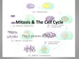
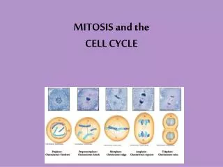
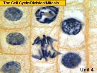
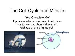
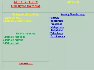
Mitosis • It is an important and specific division of the mother cell into TWO cells identical to the mother cell. • This cell division plays an important role in the growth • We can divide mitosis into five stages
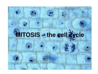
Interphase • The G1, S, and G2 phases, are very important for the successful completion of mitosis. • During G1, cells undergo the major portion of their growth. • During the S phase, each chromosome replicates to produce two sister chromatids, which remain attached to each other at the centromere. • After the chromosomes have replicated in S phase, they remain fully extended and uncoiled. This makes them invisible under the light microscope. • In G2 phase, they begin the long process of condensation, coiling ever more tightly.
Prophase • When the chromosome condensation initiated in G2 phase reaches the point at which individual condensed chromosomes first become visible with the light microscope. • The condensation process continues throughout prophase; consequently, some chromosomes that start prophase as minute threads appear quite bulky before its conclusion. • The assembly of the microtubular apparatus that will later separate the sister chromatids also continues during prophase. • In animal cells, the two centriole pairs formed during G2 phase begin to move apart early in prophase, forming between them an axis of microtubules referred to as spindle fibers.
By the time the centrioles reach the opposite poles of the cell, they have established a bridge of microtubules called the spindle apparatus between them. • During the formation of the spindle apparatus, the nuclear envelope breaks down and the endoplasmic reticulum reabsorbs its components. • At this point, then, the microtubular spindle fibers extend completely across the cell, from one pole to the other. • Their orientation determines the plane in which the cell will subsequently divide, through the center of the cell at right angles to the spindle apparatus.
Metaphase • The second stage of mitosis, metaphase, is the phase where the chromosomes align in the center of the cell. • When viewed with a light microscope, the chromosomes appear to array themselves in a circle along the inner circumference of the cell, as the equator girdles the earth. • An imaginary plane perpendicular to the axis of the spindle that passes through this circle is called the metaphase plate. • Positioned by the microtubules attached to the kinetochores of their centromeres, all of the chromosomes line up on the metaphase plate.
Anaphase and Telophase: • The centromeres of all the chromosomes separate simultaneously, but the mechanism that achieves this synchrony is not known. • In the process, two forms of movement take place simultaneously, each driven by microtubules. • First, the poles move apart as microtubular spindle fibers physically anchored to opposite poles slide past each other, away from the center of the cell. • Second, the centromeres move toward the poles as the microtubules that connect them to the poles shorten.
When the sister chromatids separate in anaphase, the accurate partitioning of the replicated genome—the essential element of mitosis—is complete. • In telophase, the spindle apparatus disassembles, as the microtubules are broken down into tubulin monomers that can be used to construct the cytoskeletons of the daughter cells. • A nuclear envelope forms around each set of sister chromatids, which can now be called chromosomes because each has its own centromere. • The chromosomes soon begin to uncoil into the more extended form that permits gene expression.
Cytokinesis • The eukaryotic cell has partitioned its replicated genome into two nuclei positioned at opposite ends of the cell. • The replication of organelles takes place before cytokinesis, often in the S or G2 phase. • Cell division is still not complete at the end of mitosis, however, because the division of the cell proper has not yet begun. • The phase of the cell cycle when the cell actually divides is called cytokinesis. • It generally involves the cleavage of the cell into roughly equal halves.
Cytokinesis in Animal Cells • In animal cells and the cells of all other eukaryotes that lack cell walls, cytokinesis is achieved by means of a constricting belt of actin filaments. • As these filaments slide past one another, the diameter of the belt decreases, pinching the cell and creating a cleavage furrow around the cell’s circumference. • As constriction proceeds, the furrow deepens until it eventually slices all the way into the centre of the cell. • At this point, the cell is divided in two.
Cytokinesis in Plant Cells • These cells assemble membrane components in their interior, at right angles to the spindle apparatus. • This expanding membrane partition, called a cell plate, continues to grow outward until it reaches the interior surface of the plasma membrane and fuses with it, effectively dividing the cell in two. • Cellulose is then laid down on the new membranes, creating two new cell walls. • The space between the daughter cells becomes impregnated with pectins and is called a middle lamella.