Download
1 / 95
1k likes | 1.5k Vues
MECHANICS OF PULMONARY VENTILLATION. by- Dr. Shafali Singh. Learning objectives. List the major muscles involved in respiration, and state the role of each. Define the basic measures of lung volume and give approximate values for each in a normal adult.

E N D
MECHANICS OF PULMONARY VENTILLATION by- Dr. Shafali Singh
Learning objectives • List the major muscles involved in respiration, and state the role of each. • Define the basic measures of lung volume and give approximate values for each in a normal adult. • Pressure changes during breathing: Intrapulmonary, Intrapleural, Transpulmonary pressures • Define compliance, and give examples of diseases in which it is abnormal. • Describe the chemical composition and function of surfactant
Mechanics of Pulmonary Ventilation • Muscles That Cause Lung Expansion and Contraction: • Normal quiet breathing by movement of the diaphragm. Inspiration-contraction of the diaphragm Expiration- elastic recoil of the lungs, chest wall, and abdominal structures compresses the lungs and expels the air
Accessory muscles Muscles that raise the rib cage are the external intercostals, but others that help are (1) sternocleidomastoid muscles (2) anterior serrati, (3) scaleni The muscles that pull the rib cage downward during expiration are mainly (1) abdominal recti(2) internal intercostals.
Mechanisms of Pulmonary Ventilation Figure 23.15a-d
Elastic behaviour of lung • Collagen and elastin fibers: “nylon stocking elasticity” • Surface tension
Lung Recoil • Represents forces that develop in the wall of the lung as the lung expands. • As the lung enlarges, recoil increases; • As the lung gets smaller, recoil decreases. • Recoil, as a force, always acts to collapse the lung.
Before Inspiration The glottis is open, and all respiratory muscles relaxed (FRC).This is the neutral or equilibrium point of the respiratory system. The intrapleural force and recoil force are equal and opposite, and because no air is flowing through the open glottis, alveolar pressure must be zero. By convention, the atmospheric pressure is set to equal zero.
During Inspiration Inspiration is induced by the contraction of the diaphragm which expands the chest cavity. The net result is to decrease (make more negative) intrapleural pressure. The expansion of the lung causes the gases in the alveoli to expand, creating a slightly negative alveolar pressure. This causes air to flow into the lung.
End of Inspiration The lung expands until the recoil force increases to equal intrapleural pressure. Once the forces are again equal and opposite, the lung is at its new larger volume. The inflowing air returns alveolar pressure toward zero, and when it reaches zero, airflow stops. Under resting conditions, about 500 ml of air flows into the lung system in order to return alveolar pressure back to zero.
Movement of Air In and Out of the Lungs and the Pressures That Cause the Movement • Pleural Pressure and Its Changes During Respiration
Q At the end of a quiet inspiration, intra-alveolar pressure is normally a. 240 cmH2O b. 24 cmH2O c. 0 cm H2O d. 14 cmH2O e. 140 cmH2O
The diagram below illustrates the change in intrapleural pressure during a single breath. At which point on the diagram is inspiratory airflow the greatest? a. A b. B c. C d. D e. E
At which point on the diagram is lung volume the greatest? a. A b. B c. C d. D e. E
Before inspiration has begun, airway pressure? IPP? • zero (no flow), -5cmH2O. • Airway pressure at end-inspiration? • zero again (no flow). • Pa and Pip during FORCED expiration? • both Pip and Pa increase significantly
During forced expiration what happens due to the P drop along the airway as flow begins? • there is compression and collapse of airway in compressible airways when the Pa falls below Pip (negative distending P). • Why does increasing the force of expiration not increase the flow during a forced expiration when there is airway collapse? • any increase in expiratory effort that increases compression on the thorax raises Pa and Pip by the SAME amt, so the driving P for flow doesnt change and flow becomes effort independent.
Collapse of the respiratory passage way during maximum expiratory effort, an effect that limits expiratory flow rate
What happens if the parietal pleura is penetrated by external trauma or the visceral pleural is interrupted by a ruptured pulmonary cyst? • Air will enter the pleural cavity and the lung will collapse by virtue of its own intrinsic elasticity • When air enters the pleural cavity, what does this cause? • Pneumothorax • A pneumothorax results in a collapsed lung on ____ side as the injury? • same • What causes the lung to collapse in a pneumothorax? • Surface tension and recoil of elastic fibers
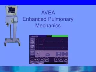
POSITIVE-PRESSURE RESPIRATION • Assisted Control Mode Ventilation (ACMV) • Positive End-Expiratory Pressure (PEEP) • ContinuousPositive Airway Pressure (CPAP)
PHYSIOLOGICAL CONDITIONS & FACTORS INFLUENCING INTRAPULMONARY PRESSURE Valsalva’smanoeuvre- 1.Blowing a balloon, 2.Sneezing 3.Defaecation, 4.Micturition 5.Coughing, 6.Parturition Muller’s manoeuvre- Suckling of fluids
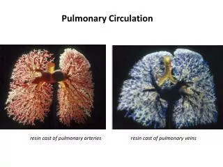
Effects of intra pleural pressure onpulmonary blood flow and volume Inspiration:Intrapleuralpressure becomes more negative (decreases). Expiration: Intrapleuralpressure becomes more positive (increases), Systemic venous return and output of the right ventricle are decreased. The return of blood and output of the left ventricle are increased, caus-ing increased systemic arterial pressure. reflex decrease in heart rate. • Systemic venous return and right ventricular output are increased. • Venous return to the left heart, and the output of the left ventricle is decreased, causing decreased systemic arterial pressure. • Reflex increase in heart rate
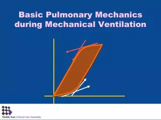
Compliance describes the distensibility of the lungs and chest wall. ■ Is inversely related to elastance, which depends on the amount of elastic tissue. ■ Is the slope of the pressure–volume curve. ■ Is the change in volume for a given change in pressure. Pressure refers to transmural, or transpulmonary, pressure (i.e., the pressure difference across pulmonary structures).
Compliance(stretchability)of the Lungs • Lung compliance is the change in lung volume (tidal volume) divided by the change in surrounding pressure • The total compliance of both lungs together is 200ml air/cm transpulmonary pressure. • Lung thorax system together is 110ml/cmH2O
Emphysema [increased lung compliance]. • In this condition, there is loss of elastic fibers in the lungs with resultant increase in compliance. The volume-pressure slope becomes steeper. The collapsing force at a given volume is decreased. • At the original value for FRC, the tendency for the lungs to collapse is less than the tendency of the lungs to expand. Thus, the combined chest wall and lung system seeks a new higher FRC to balance the opposing forces. • A patient with emphysema is said to breathe at higher lung volumes and will have a barrel-shaped chest.
Fibrosis [decreased lung compliance] • In this condition, there is stiffening of lung tissues and decreased compliance i.e. decreased slope of the curve for the lung. • At the original FRC, the tendency of the lungs to collapse is more than the tendency of the chest wall to expand with loss of balance. To reinstall balance, the chest wall and lung system seeks a new lower FRC.
Factors affecting only the lung compliance • Lung volume • Phase of respiratory cycle • Effect of gravity • Surface tension
Surface tension • Surface tension forces are created whenever there is a liquid–air interface. • The molecules of water attract each other much more than the air above them • Feature of ST is that in a Curved surface it develops a pressure
The relationship between the surface tension and the pressure inside a bubble is given by the law of LaPlace. • In the alveoli, they act to collapse the alveoli; therefore, these forces contribute to lung recoil.