Download
1 / 98
1.01k likes | 1.29k Vues
Functional anatomy of pulmonary system, pulmonary circulation and mechanics of breathing. Presenter: Dr. Satyajit Majhi Moderator: Dr. J.P. S harma. University College of Medical Sciences & GTB Hospital, Delhi. Email: anaesthesia.co.in@gmail.com. www.anaesthesia.co.in.

E N D
Functional anatomy of pulmonary system, pulmonary circulation and mechanics of breathing Presenter: Dr. Satyajit Majhi Moderator: Dr. J.P. Sharma University College of Medical Sciences & GTB Hospital, Delhi Email: anaesthesia.co.in@gmail.com www.anaesthesia.co.in
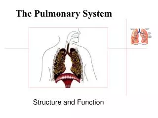
5 Functions of the Respiratory System • Provides extensive gas exchange surface area between air and circulating blood • Moves air to and from exchange surfaces of lungs • Protects respiratory surfaces from outside environment • Produces sounds • Participates in olfactory sense
The Nose • Air enters the respiratory system: • through nostrils or external nares • into nasal vestibule • Nasal hairs: • are in nasal vestibule • are the first particle filtration system
The Nasal Cavity • The nasal septum: • divides nasal cavity into left and right • Superior portion of nasal cavity is the olfactory region: – provides sense of smell • Mucous secretions from par nasal sinus and goblet cells: • clean and moisten the nasal cavity • Lined by ciliated mucosal layer
Epistaxis • Most common site Little’s area • Situated anterior inferior part of nasal septum. • Anastomosis of 4 arteries, anterior ethmoidal, septal branch of superior labial, septal branch of sphenopalatine and greater palatine. • Woodruff area, anastomosis of sphenopalatine artery and posterior pharyngeal artery causes posterior epistaxis
Air Flow Meatuses • Constricted passageways that produce air turbulence: • warm and humidify incoming air • trap particles • During exhalation these structures: • Reclaim heat and moisture • Minimize heat and moisture loss
The Palates • Hard palate: • forms floor of nasal cavity • separates nasal and oral cavities • Soft palate: • extends posterior to hard palate • divides superior nasopharynx from lower pharynx
The Pharynx and Divisions • A chamber shared by digestive and respiratory systems • Extends from internal nares to entrances to larynx and esophagus • Nasopharynx • Oropharynx • Laryngopharynx
The Nasopharynx • Superior portion of the pharynx • Contains pharyngeal tonsils and openings to left and right auditory tube • Pseudo-stratified columnar epithelium The Oropharynx • Middle portion of the pharynx • Communicates with oral cavity • Stratified squamous epithelium The Laryngopharynx • Inferior portion of the pharynx • Extends from hyoid bone to entrance to larynx and esophagus
Cartilages of the Larynx • Air flow from the pharynx, enters the larynx: • a cartilaginous structure that surrounds the glottis • 3 large, unpaired cartilages form the larynx: • the thyroid cartilage • the cricoid cartilage • the epiglottis
The Thyroid Cartilage • Also called the Adam’s apple • Is a hyaline cartilage • Forms anterior and lateral walls of larynx • Ligaments attach to hyoid bone, epiglottis, and laryngeal cartilages
The Cricoid Cartilage • Is a hyaline cartilage • Form posterior portion of larynx • Ligaments attach to first tracheal cartilage • Articulates with arytenoid cartilages The Epiglottis • Composed of elastic cartilage • Ligaments attach to thyroid cartilage and hyoid bone
Cartilage Functions • Thyroid and cricoid cartilages support and protect: • the glottis • the entrance to trachea • During swallowing: • the larynx is elevated • the epiglottis folds back over glottis • Prevents entry of food and liquids into respiratory tract
Cartilage Functions 3pairs of Small Hyaline Cartilages of the Larynx arytenoid cartilages, corniculate (Santorini) cartilages and Cuneiform (Wrisberg) cartilages • Corniculate and arytenoid cartilages function in: • opening and closing of glottis • production of sound
Ligaments of the Larynx • Vestibular ligaments and vocal ligaments: • extend between thyroid cartilage and arytenoid cartilages • are covered by folds of laryngeal epithelium that project into glottis 1) The Vestibular Ligaments • Lie within vestibular folds: • which protect delicate vocal folds
Speech • Speech – intermittent release of expired air while opening and closing the glottis • Pitch – determined by the length and tension of the vocal cords • Loudness – depends upon the force at which the air rushes across the vocal cords • The pharynx resonates, amplifies, and enhances sound quality • Sound is “shaped” into language by action of the pharynx, tongue, soft palate, and lips
The Laryngeal Musculature • Laryngeal muscle can be – Extrinsic muscles that • Elevates or depresses the hyoid bone • Intrinsic muscles that: • control vocal folds • open and close glottis • Coughing reflex: food or liquids went “down the wrong pipe”
Nerve supply of Larynx • Mucous membrane above vocal fold – internal laryngeal branch of superior laryngeal branch of vagus nerve • Below that its supplied by – recurrent laryngeal nerve (RLN) • All intrinsic muscle, except cricothyroid – RLN, cricothyroid by external laryngeal branch of SLN
Laryngeal paralysis RLN SLN COMBINED
Sphincter Functions of the Larynx • The larynx is closed during coughing, sneezing, and Valsalva’s maneuver • Valsalva’s maneuver • Air is temporarily held in the lower respiratory tract by closing the glottis • Causes intra-abdominal pressure to rise when abdominal muscles contract • Helps to empty the rectum • Acts as a splint to stabilize the trunk when lifting heavy loads
Organization of the Respiratory System • The respiratory system is divided into the upper respiratory system, above the larynx, and the lower respiratory system, from the larynx down
The Respiratory Tract • Consists of a conducting portion: • from nasal cavity to terminal bronchioles • Transitional portion –the respiratory bronchioles and alveolar ducts • Respiratory portion: • the alveoli and alveolar sac Alveoli • Are air-filled pockets within the lungs • where all gas exchange takes place
The Trachea • Extends from the cricoid cartilage into mediastinum • Formed of rings of cartilages, incomplete posteriorly • Lined by ciliated columnar epithelium • It bifurcates into right and left main bronchi at the level of T5
The Tracheal Cartilages • 15–20 tracheal cartilages: • strengthen and protect airway • discontinuous where trachea contacts esophagus • Ends of each tracheal cartilage are connected by: • an elastic ligament and trachealis muscle
The Primary Bronchi • Right and left primary bronchi: • separated by an internal ridge (the carina) The Right Primary Bronchus • Is larger in diameter and shorter (2.5 cm) than the left • Descends at a steeper angle (25⁰) – The Left Primary Bronchus • Is narrower and longer (5cm) • Descends at broader angle (55⁰)
Bronchi subdivide into secondary bronchi, each supplying a lobe of the lungs • Air passages undergo 23 orders of branching in the lungs • Tissue walls of bronchi mimic that of the trachea • As conducting tubes become smaller, structural changes occur • Cartilage support structures change • Epithelium types change • Amount of smooth muscle increases
Secondary Bronchi • Branch to form tertiary bronchi, also called the segmental bronchi • Each segmental bronchus: • Supplies air to a single bronchopulmonary segment • The right lung has 10 • The left lung has 8 or 9
Bronchial Structure • The walls of primary, secondary, and tertiary bronchi: • contain progressively less cartilage and more smooth muscle • increasing muscular effects on airway constriction and resistance
The Bronchioles • Each tertiary bronchus branches into multiple bronchioles • 1 tertiary bronchus forms about 6500 terminal bronchioles • Bronchioles branch into terminal bronchioles
Bronchiole Structure • Bronchioles: • have no cartilage • are dominated by smooth muscle Autonomic Control • Regulates smooth muscle: • controls diameter of bronchioles • controls airflow and resistance in lungs
Bronchodilation • Dilatation of bronchial airways • Caused by sympathetic ANS activation • Reduces resistance Bronchoconstriction • Constricts bronchi • Caused by: • parasympathetic ANS activation • histamine release (allergic reactions)
Pulmonary Lobules • Are the smallest compartments of the lung • Are divided by the smallest trabecular partitions (interlobular septa) • Each terminal bronchiole delivers air to a single pulmonary lobule • Each pulmonary lobule is supplied by pulmonary arteries and veins
Exchange Surfaces • Within the lobule: • each terminal bronchiole branches to form several respiratory bronchioles, where gas exchange takes place
Alveolar Organization • Respiratory bronchioles are connected to alveoli along alveolar ducts • Alveolar ducts end at alveolar sacs: • common chamber connected to many individual alveoli
An Alveolus • Has an extensive network of capillaries • Is surrounded by elastic fibers Alveolar Epithelium • Consists of simple squamous epithelium • Consists of thin, delicate Type I cells • Patrolled by alveolar macrophages, also called dust cells • Contains septal cells (Type II cells) that produce Surfactant- an oily secretion which • Contains phospholipids and proteins • Coats alveolar surfaces and reduces surface tension
Respiratory Membrane - The thin membrane of alveoli where gas exchange takes place 3 Parts of the Respiratory Membrane • Squamous epithelial lining of alveolus • Endothelial cells lining an adjacent capillary • Fused basal laminae between alveolar and endothelial cells Diffusion- Across respiratory membrane is very rapid: • because distance is small • gases (O2 and CO2) are lipid soluble
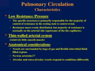
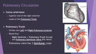
Blood Supply to Respiratory Surfaces • Each lobule receives an arteriole and a venule • respiratory exchange surfaces receive blood: • from arteries of pulmonary circuit • a capillary network surrounds each alveolus: • as part of the respiratory membrane • blood from alveolar capillaries: • passes through pulmonary venules and veins • returns to left atrium
Gross Anatomy of the Lungs • Left and right lungs: • are in left and right pleural cavities • The base: • inferior portion of each lung rests on superior surface of diaphragm