Pulmonary System
Pulmonary System. Essentials of Exercise Physiology. Respiration. External respiration: ventilation and exchange of gasses in the lungs (pulmonary function). Internal respiration : ventilation and exchange of gasses in the tissues (pulmonary function). Functions of Respiratory System.

Pulmonary System
E N D
Presentation Transcript
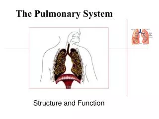
Pulmonary System Essentials of Exercise Physiology
Respiration • External respiration:ventilation and exchange of gasses in the lungs (pulmonary function). • Internal respiration: ventilation and exchange of gasses in the tissues (pulmonary function).
Functions of Respiratory System Primary purpose of respiratory system is: Provide means of oxygen exchange between external environment and body Provide a means of carbon dioxide exchange between the body and the external environment Exchange occurs as result: Ventilation: mechanical Diffusion: random movement
Functions of Respiratory System Respiratory system also helps regulate acid-base balance in body, especially during exercise. Cl-+ H+ + NaHCO3 NaCl + H2CO3 CO2 + H2O
Acid - Base Balance • Acids - molecules which can liberate hydrogen ions • Bases - molecules which can accept hydrogen ions • Buffer - resists changes in pH by either accepting hydrogen ions or liberating them depending upon local conditions
Structure Pulmonary System • Right and left lungs enclosed by membranes called pleura • Visceral pleura adheres to outer surface of lungs • Parietal pleura adheres to thoracic wall and diaphragm
Intrapleural Space • Contains fluid which lubricates pleura • Creates a low pressure area • pressure is below atmospheric during inspiration, allowing the lungs to inflate
Functional Zones of Air Passages • Conducting zone • passageways leading to respiratory zone • area where no gas exchange occurs • nasal cavity, pharynx, larynx, trachea, bronchioles • Respiratory zone • where gas exchange actually occurs • alveoli
Roles of Conducting Zone • Warms air • Mucus traps small particles • Cilia sweep particles upwards • Macrophages engulf foreign particles
Roles of Respiratory Zone • Provides large surface area for gas exchange • 600 million alveoli • Total surface area is 60 – 80 square meters or about size of half a tennis court • Provides a very thin barrier for gas exchange • 2 cell layers thick
Alveoli • Type II alveolar cells secrete pulmonary surfactant • form a monomolecular layer over alveolar surfaces • surfactant stabilizes alveolar volume by reducing surface tension created by moisture
Mechanics of Ventilation • Change in thoracic cavity volume produces corresponding change in lung volume • Increase in lung volume results in decrease in lung pressure (Boyle’s law) • Differences in pressure pulls air into the lungs • pressure within the lungs becomes less than the atmospheric pressure • bulk flow (from high pressure to low pressure)
Muscles of Inspiration • Diaphragm • contracts, flattens, & moves downward up to 10 cm • enlarges & elongates chest cavity, expands volume • during quiet breathing diaphragm works alone • External intercostals, pectoralis minor, sternocleidomastoid & scaleni • lift ribs up and outwards • during exercise, accessory muscles called into play
Muscles of Expiration • Expiration during quiet breathing is passive due to elastic recoil of chest cavity • Decrease in lung volume forces air out of lungs • During exercise and voluntary hyperventilation, • rectus abdominus, transverse abdominus: push diaphragm up • internal intercostals: pull ribs downwards
Total Lung Capacity • Tidal volume (VT) • amount either inspired or expired during normal ventilation • Inspiratory reserve volume • maximal volume inspired after a normal inspiration • Expiratory reserve volume • volume expired after a normal expiration • During exercise VT increases largely from IRV. • Residual volume • volume remaining in lungs after maximal expiration
Lung Capacities • Total lung capacity • volume within lung after a maximal inspiration • Inspiratory capacity • maximal volume inspired from the end of tidal expiration • Functional residual capacity • volume in lungs after normal expiration • Vital capacity • maximal volume expired after maximal inspiration
Dynamic Lung Volumes • Depend on volume and speed of air movement; more useful in diagnosing lung disease. • FEV: Forced Expiratory Volume. Volume that can be forcefully expired after maximal inspiration within given time, usually 1 sec. • MVV: Maximal Voluntary Ventilation. Volume of air that can be ventilated by maximal effort in one minute. Breathe maximally for 12 (or 15) seconds and total volume recorded, multiplied by five (or 4).
Minute Ventilation • Volume of gas ventilated in one minute • equal to tidal volume times frequency • Rest in 70 kg man, 6.0 L/min = 0.5 L x 12 • Maximal exercise, 120-175 L/m = 3-3.5 x 40-50 • increases as oxygen consumption increases • closely associated with CO2 production ERROR
Anatomical vs Physiological Dead Space • Anatomical dead space • areas of conducting zone not designed for diffusion of gases • VT = VA + VD • At rest, VT = 500 ml = 350 ml + 150 ml • Physiological dead space • areas of lung and pulmonary capillary bed which are unable to perform gas exchange as designed
Physiologic Dead Space • Optimal diffusion requires matching of ventilation to perfusion: 1 ventilated alveoli/ 1 blood perfused alveoli • Ventilation (V) / perfusion (Q) is not equal across the lung • Top of lung is poorly perfused • V / Q = 3.3 at top of lung • Bottom of lung has more perfusion than ventilation • V / Q = .63 at bottom of lung • V / Q values above .5 are generally adequate
Minute Ventilation in Exercise • Adjustments in breathing rate and depth maintain alveolar ventilation as exercise. • Trained athletes maintain alveolar ventilation by increasing VT and minimal increase rate. • Deeper breathing causes a greater percentage of incoming “fresh” VT to enter alveoli. • Increasing VT in exercise results from encroaching primarily on IRV or ERV? • VT plateaus at about 60% vital capacity.
Disruptions in Normal Breathing • Dyspnea shortness of breath or subjective distress in breathing. • Hyperventilation≠ Hyperpnea • Valsalva maneuver: forced exhalation against closed glottis. What happens to blood pressure?
Gas Exchange Fick’s Law • Diffusion occurs at a rate which is proportional to differences in partial pressure and the surface area available and is inversely proportional to the thickness of the membrane. • Diffusion rate = (P1 - P2) area thickness