The Pulmonary System
The pulmonary system comprises the lungs, which weigh around 1 kg and can hold 4-6 liters of air. Lung tissue covers an area equivalent to half a tennis court, significantly larger than the external body surface. Key functions include ventilation (breathing), air conduction, and gas exchange (O2 and CO2). The system features critical structures like the nasal cavity, larynx, trachea, bronchi, and alveoli, where 300 million alveoli exchange gases efficiently. Mechanics of breathing involve pleura membranes and pressure changes during inspiration and expiration.

The Pulmonary System
E N D
Presentation Transcript
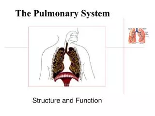
The Pulmonary System Structure and Function
Lungs • Lung tissue weighs 1 kg and covers half a tennis court (50-100 square feet) • Lung tissue is 20-50 times larger than the body’s external surface • Hold 4-6 liters of air. • Unattached to ribs; suspended inside the pleural sacs.
Function • Ventilation. Breathing (air in and air out) • Conduction. • Movement of air through the pulmonary system • Respiration. • Gas exchange (O2 and CO2)
Conduction Zone (humidify, filter) • Nasal cavity and Pharynx • Nose moistens, warms, and filters air; mouth does not. • Larynx - voice box • Epiglottis • Valsalva maneuver
Conduction Zone (humidify, filter) • Trachea • conducting tube (transports air) • Bronchi • Branches • contains muscle, serves to dilate and constrict • Anatomic dead space
Respiration • Bronchioles • further branching • Alveolar sacs (300 million) • Each alveoli is surrounded by a network or covering of capillaries. • Almost forms a “sheet” of blood. • At rest, a single blood cell passes by 2 or 3 alveoli in about 0.5 to 1.0 seconds
Respiration (gas exchange) • Occurs through thin walls (0.3 micrometers) • Diffusion of gases from high to low concentration.
Types of Respiration • Pulmonary (external) • Transfer of O2 and CO2 at the lungs. • 250 mL of oxygen is exchanged per minute at rest • 200 mL of CO2 is exchanged per minute at rest • These numbers can increase up to 25 times during heavy exercise • Cellular (internal) respiration - transfer of O2 and CO2 in the tissues.
Pleura • Pair of membranes (inner and outer) surrounding the lungs • Fluid in between two sacs provides the only attachment of the lungs to the thorasic cavity (ribs).
Pressure • Inspiration • Air moves into the lungs due to a lower pressure inside the lungs • Expansion of the rib cage and the lowering of the diaphragm increase the volume • As the volume gets larger, the pressure becomes lower.
Pressure • Expiration • Air moves out of the lungs due to a higher pressure inside the lungs • Constriction of the rib cage and the raising of the diaphragm decrease the volume • As the volume gets smaller, the pressure becomes higher.
Muscles of Ventilation • Inspiration • Rest • diaphragm and E.IC muscles • Exercise • pectorals, scalenes, SCM • Expiration • Rest • no muscles • Exercise • abs, I.IC. muscles)
Lungs and Exercise • At rest, the blood is 97-98% saturated with oxygen • Exercise, the blood remains 97-98% saturated • Healthy lungs do not limit a person’s ability to exercise • However, respiratory muscles need to be trained like any other skeletal muscle