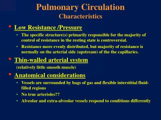
Pulmonary Circulation
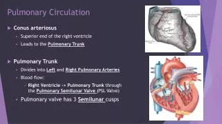
Pulmonary Circulation. Conus arteriosus Superior end of the right ventricle Leads to the Pulmonary Trunk Pulmonary Trunk Divides into Left and Right Pulmonary Arteries Blood flow: Right Ventricle -> Pulmonary Trunk through the Pulmonary S emilunar Valve (PSL Valve)

Pulmonary Circulation
E N D
Presentation Transcript
Pulmonary Circulation • Conusarteriosus • Superior end of the right ventricle • Leads to the Pulmonary Trunk • Pulmonary Trunk • Divides into Left and Right Pulmonary Arteries • Blood flow: • Right Ventricle -> Pulmonary Trunk through the Pulmonary Semilunar Valve (PSL Valve) • Pulmonary valve has 3 Semilunar cusps
Systemic Circulation • Blood leaves Left Ventricle through … • Aortic Semilunar Valve (ASL Valve) into Ascending Aorta • Ascending Aorta turns to the left into the … • Aortic arch and becomes the …. • Descending Aorta
Systemic Circulation • Structural Differences between the Left & the Right Ventricle: • Left Ventricle • Round • Thicker myocardium wall -> more pressure • Right Ventricle • Pouch shaped • Thinner myocardium wall -> less pressure
Coronary Sinus • Cardiac veins return blood to coronary sinus • Cardiac sinus opens into Right Atrium
Writing Activity (10 points) • You are a Erythrocyte (RBC) traveling through the human body. Describe the journey! • Condition in lungs for O2 pick up • Heart: atria, ventricles, 4 valves • Systemic pathway • Pulmonary pathway • Conditions of tissue for O2 dispersal & CO2 pick up • 2 Bonus points: if all possible names for valves are given • Example: Atrioventricular valve, Mitral valve, …
Foramen Ovale • Before Birth; an opening through the Interatrial Septum • Connects the two Atria • Eliminates blood being sent to the lungs in a Fetus!! • Seals off at birth -> Fossa Ovalis • Blue Baby • Foramen ovale does not deal off after birth • Requires surgery
Heart Valves Tricuspid Valve Bicuspid Valve Mitral Valve Pulmonary Semilunar Valve Left AV valve: Bicuspid valve Mitral valve Right AV Valve: Tricuspid Aortic Semilunar Valve Pulmonary Semilunar Valve Aortic Semilunar Valve
Cardiac Conduction • Heart Beat • A single contraction of the heart • The entire heart contracts in sequence • Atria • Ventricles
Cardiac Conduction • Structures of the Conducting System • Sinoatrial (SA) nodes • In the wall of the Right Atrium • Atrioventricular (AV) node • Between Atrium & Ventricle • Conducting Cells • Throughout myocardium
The Cardiac Cycle • Begins at the Sinoatrial ( SA) Node with an Action Potential • The Action Potential is transmitted through the conducting System: • SA node • AV node • Produces an Action Potential in the Cardiac muscle cells • Contractile cells
5 Steps to the Cardiac Cycle: • Ventricular diastole • Atrial systole begins • Atrial diastole • Ventricular systole 1st phase • Ventricular systole 2nd phase Diastole = Relaxation Systole = Contraction
5 Steps to the Cardiac Cycle: • Ventricular diastole • All chambers are relaxed Diastole = Relaxation Systole = Contraction
5 Steps to the Cardiac Cycle: • Atrial systole begins • Atria contract & force blood into ventricles Diastole = Relaxation Systole = Contraction
5 Steps to the Cardiac Cycle: • Atrial diastole • Atrium contraction ends • Atrium relaxes • Ventricle contractions begin Diastole = Relaxation Systole = Contraction
5 Steps to the Cardiac Cycle: • Ventricular systole 1st phase • Contraction pushes AV valves close Diastole = Relaxation Systole = Contraction
5 Steps to the Cardiac Cycle: • Ventricular systole 2nd phase • Increase in pressure forces SL valves to open • Blood is ejected Diastole = Relaxation Systole = Contraction
5 Steps to the Cardiac Cycle: • Ventricular diastole • Atrial systole begins • Atrial diastole • Ventricular systole 1st phase • Ventricular systole 2nd phase Diastole = Relaxation Systole = Contraction
Sinoatrial (SA) Node • Pacemaker of the heart • Initiates heart beak • Sends excitatory impulses every 0.85 seconds • Contracts: • Atrium • Impulse is send to AV node
Atrioventricular (AV) Node • Sends impulses to AV bundles • Bundles branch into Purkinje fibers • Cause ventricle to contract
Cardiac Cycle - Review • Is the period between the start of one heartbeat and the beginning of the next • Includes contraction & relaxation with the Atria & Ventricle • Systole • Contraction • Diastole • relaxation
Phases of the Cardiac Cycle Atrial systole Atrial diastole Ventricular systole phase 1 Ventricular diastole Ventricular systole phase 2
Cardio dynamics Vagus Nerve (Cranial Nerve X) carries fibers to the heart • Cardiac center is in the Medulla Oblongata • Cardio acceleration is controlled by the sympathetic neurons • Cardio inhibition is controlled by the parasympathetic neurons