Download
1 / 34
390 likes | 925 Vues
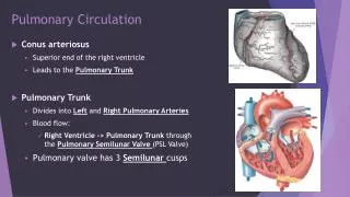
Diseases of the Pulmonary Circulation. J.B. Handler, M.D. University of New England Physician Assistant Program. PE- pulmonary embolus DVT- deep vein thrombophlebitis CO- cardiac output HF- heart failure OCP- oral contraceptive pill PAP- pulmonary artery pressure

E N D
Diseases of the Pulmonary Circulation J.B. Handler, M.D. University of New England Physician Assistant Program
PE- pulmonary embolus DVT- deep vein thrombophlebitis CO- cardiac output HF- heart failure OCP- oral contraceptive pill PAP- pulmonary artery pressure V/Q- ventilation/perfusion CVP- central venous pressure RR- respiratory rate P2- pulmonic valve component of the 2nd heart sound (S2) Vit-vitamin Dx- diagnosis INR- international normalized ratio S4- abnormal 4th heart sound NSST-T- non specific ST-T wave changes ST- sinus tachycardia ABG- arterial blood gas PuVR- pulmonary vascular resistance PPH- primary pulmonary hypertension RAE- right atrial enlargement RVE- right ventricular enlargement S2- second heart sound Rx- treatment PTT- partial thromboplastin time Abbreviations
Pulmonary Thromboembolism • Embolization of thrombus from the venous system into the pulmonary arterial circulation; common, serious and life threatening • 200,000 annual mortality in USA • 3rd leading cause of in-hospital deaths • Majority of deaths from PE unrecognized until post-mortem • 5-7% mortality rate of diagnosed cases with a 40-50% mortality rate of undiagnosed cases
Pulmonary Embolism • The presence of DVT should always sound an alarm. Prior DVT or PE carry a substantial risk for subsequent PE. • Symptoms: Often very difficult to recognize as symptoms are not specific to PE. • 97% of patients with PE will have at least one of the following: tachypnea, dyspnea, pleuritic chest pain (infers infarction). About 1/3 of patients will have tachycardia.
DVT Images.google.com
Pulmonary Thromboembolism • DVT is the most common site of development of thrombus. Usually popliteal and/or iliofemoral vein involvement with subsequent embolization (50-60%) to the pulmonary circulation. • Calf involvement alone carries much less risk of significant embolism. • Calf vein propagation to popliteal and iliofemoral veins occurs in 20% of patients with initial calf involvement. • Pelvic vein thrombosis also common source. • 50% cases of PE lack characteristic symptoms; essential to have high index of suspicion.
Risk Factors for DVT • Venous stasis: Immobility, post-op (large operations, orthopedic procedures), post stroke. • Increased CVP: Decreased CO, CHF, pregnancy. • Hypercoagulability: Meds (OCP), diseases (malignancy), and inherited- several forms: • Factor V Leiden: A hereditary hypercoagulability disorder; common (3% heterozygous incidence) in healthy adults. 20-30% of patients with DVT have this disorder. • Factor V Leiden: Modified clotting factor V, resists degradation by activated Protein Ccoagulation.
Intracardiac Pressures 4-12 4-12 4-12 4-12 4-12 4-12 Images.google.com
Pathophysiology • Obstruction to pulmonary vascular bed by thrombus. • Vasoconstriction develops in the pulmonary arteriolar bed (PuVR) as a result of: • Neurohumoral reflexes, hypoxia, and tissue injury • Result isPAP. • PAPRt heart strain CO (RVH if PAP s over time) RV failure: CorPulmonale. • Impaired gas exchange: alveolar dead spacefrom vascular obstruction.
Pathophysiology • physiologic dead space, CO, hypoxemia and surfactant depletion result in atelectasis. • Atelectasis contributes to shunting (below). • Right to left shunting plays a small role: • Atelectasis. • Large PE: Redirection of blood flow into “normal” lung but if inadequate alveolar reserveshunting. • Loss of surfactant, edema and hemorrhage lead to decreased pulmonary compliance, work of breathing.
Ventilation/Perfusion Images.google.com
Symptoms • Dependent on number/size of the emboli and pre-existing pulmonary status. • Constellation of findings: dyspnea, pleuritic (inspiratory) chest pain and tachypnea are predominant symptoms/signs; others include palpitations, wheezing and hemoptysis. • Note: must have high index of suspicion in order to make Dx in many patients.
Signs • Signs: Increased RR, tachycardia, crackles, S4 gallop, decreased S2 splitting with increased P2; friction rubs and cyanosis less common. • Clinical findings of DVT present in only 30-40% of patients with PE but 60-70% will have evidence of DVT by non-invasive imaging (see below).
Investigational Findings • ECG changes common: ST, NSST-T changes. • ABG: Respiratory alkalosis, decreased PO2. • Plasma D-dimer: (>300-500ng/ml) a degradation product of cross-linked fibrin- usually elevated in presence of thrombus (97% sensitivity), but not specific (45%). Commonly combined with other diagnostic studies (CT angiography)predicts likelihood of PE. • CxR: often abnormal- atelectasis, pleural effusions, pleural based infiltrates; necessary to exclude other pulmonary pathology.
V/Q Lung Scan • Radioisotopes used to image ventilation and perfusion. Defects develop in areas of lung that remain ventilated without perfusion; inaccurate in presence of other pulmonary pathology. • Graded low (normal), intermediate and high probability for PE. • Supplanted as diagnostic test by Helical CT angiography (below).
Pulmonary Circulation Imaging • Helical (spiral) CT Angiography: Sensitive for proximal vessel thromboembolism; overall 83% sensitive, >90% specific; alternative to V/Q scanning as a screening procedure. More often done compared to V/Q. • Pulmonary angiography: reference standard for the Dx of PE- Invasive with 5% complication rate (contrast reaction, renal failure, arrhythmias) high sensitivity, specifity. Indicated when V/Q or CT scans are indeterminate, DVT studies are negative, and PE still suspected clinically.
DVT Studies • If Dx is uncertain and DVT is proven, likely that PE is present if clinically suspected. • Venous ultrasound with doppler: Non- invasive test of choice for DVT. • Contrast Venography: “reference standard”-highly accurate and invasive, has been replaced by ultrasound which is non-invasive and easier to perform.
Clinical feature Points Clinical symptoms of DVT 3 Other diagnosis less likely than PE 3 Heart rate greater than 100 beats per minute 1.5 Immobilization or surgery within past 4 weeks 1.5 Previous DVT or PE 1.5 Hemoptysis 1 Malignancy 1 Total points Integrated Approach for Dx of PE: Clinical Features + Diagnostic Testing Wells et. al
Clinical Probability of PE PE Unlikely PE Likely D-dimer assay Helical CT-PA Negative Positive Normal Findings of PE PE Excluded Rx for PE PE excluded - study Indeterminate Current fig 9-1 LE US or Pulm angiogram + study
Treatment of 1st PE • Full anticoagulation for minimum 6 months or longer depending on risk factors for recurrence. • Unfractionated heparin: Binds to and potentiates antithrombin III inactivates clotting factors IXa, Xa, XIa, XIIa; retards additional thrombus formation. • Administered by continuous IV infusion and requires frequent monitoring (highly protein bound) of PTT for dose adjustment; for 5-7 days. • Wt. Based dosing: 80U/kg loading dose, then 18U/kg/hrgoal PTT 1.5-2.5x control (46-70 sec) • Risks: Bleeding, thrombocytopenia.
Low Molecular Weight Heparin: depolymerized heparin, less protein and cellular bound, increased bioavailability; also given for 7 days. More predictive dose response, as/more effective than unfractionated heparin. Decreased risk of hemorrhage and thrombocytopenia; 1-2x daily dosing (weight based) without need for lab testing. • Warfarin- oral anticoagulant, blocks action of Vit K, inhibiting hepatic synthesis of Vit K dependent clotting factors (II, VII, IX, X). Initiated over 5-7 days (with patient on heparin) and continued for desired period of time. Goal: INR (adjusted PT) of 2-3.
Thrombolytic Therapy • Streptokinase, Urokinase and t-PA: directly lyse intravascular thrombi and accelerate resolution of emboli within 1st 24 hours compared to standard heparin therapy, but do not decrease mortality. • Indications: patients with massive PE at high risk of death (unstable on heparin). • Contraindications: active internal bleeding, stroke in prior two months, surgery or trauma in prior 6 weeks and uncontrolled hypertension.
Surgical Interventions • Mechanical or surgical thrombus extraction- last resort in a dying patient. • Vena caval interruption: Indicated in patients with DVT/PE that cannot be anticoagulated or recurrent PE while already fully anticoagulated. Vena caval filter/umbrella can be introduced percutaneously via internal jugular vein. • Prognosis of PE if diagnosed: < 3% incidence of death from recurrent PE if initial event is recognized and adequately treated.
Prevention of PE • Identify patients at high risk for DVT: lengthy hospitalization/immobilization, orthopedic surgery, esp. hip repair (incidence of DVT 10-20% if untreated with prophylactic anticoagulation and other measures). • Strategies for prevention:-early ambulation-elastic stockings-mechanical compression devices-anticoagulation.
Pharmacologic Prophylaxis • Patients with high risk for DVT and PE • Low doses (below full anticoagulation dose) of anticoagulants (’s risk of bleeding) decreases incidence of DVT/PE: Unfractionated Heparin Low molecular weight (LMW) heparin • Sometimes continued as OP (warfarin/coumadin) in patient likely to remain immobile.
Pulmonary Hypertension • Pulmonary circulation is a low pressure system with high blood flow and low PuVR (200 resistance units compared with 800-1000 RU for the systemic circulation. • In a variety of disease states (e.g. PE) there is constriction of smooth muscle in the walls of pulmonary arteriolar resistance vessels. • Pulmonary Hypertension is present when the PAP rises to a high level, inappropriate for a given level of cardiac output. • PPH- rare idiopathic disorder in woman.
Idiopathic (Primary) Pulmonary Hypertension • No other underlying cardiopulmonary disease: An arteriopathy. Medial hypertrophy, fibrosis and recanalized thrombi are common pathology findings; poor prognosis- mean survival post Dx is 2-5 years. • Marked PuVR and PAP. • Association: anorexigens, collagen-vascular disease. • Fenflouramine appetite suppressants: Marked recent in PPH (U.S. and Europe); similar events in 1960’s with Amrinox. Both taken off market.
Secondary Pulmonary HTN • Reduction of x-sectional area of the pulmonary vascular bed: • Vasoconstriction: Hypoxia from any cause (most important and potent stimulus), lactic acidosis. • Loss of vessels: Vasculitis, emphysema, interstitial dis. • Obstruction of vessels: Multiple or recurrent PE. • Chronically increased pulmonary venous pressure (HF, mitral stenosis). • pulmonary blood flow: Congenital Ht Disease. • Once present, secondary structural changes occur resulting in further destruction of vessels.
Signs and Symptoms • Dyspnea- initially exertional, then rest. • Retrosternal chest pain (mimics angina); fatigue and syncope result from decreased CO. • Physical Exam: Narrowly split or single S2, P2. • CxR: Dilated main PA with “pruning” of vessels. • Echo-Doppler: best non-invasive tool: RAE, RVE; elevated PA and RV systolic pressure. • Right heart cath: precise measurement of right heart pressures essential to Dx and Rx.
Treatment • Treat underlying disease process if present. • Supplemental chronic O2 if hypoxemic. • Full anticoagulation: PPH only • Prostacyline (Epoprostenol, Treprostinil): via continuous IV infusion potent pulmonary vasodilator; symptoms and mortality.
Treatment • Bosentan (Tracleer): new oral endothelin-1 receptor antagonist for chronic PAH; symptoms. • Sildenafil: inhibits phosphodiesterase type 5; pulmonary vasodilator- improves symptoms. • Lung or heart/lung transplant: 50-65% 2 year survival. Better long term survival with bilateral (vs. unilateral) lung transplant.