Download
1 / 67
720 likes | 828 Vues
Explore the cardiovascular system's double-pump heart, circulation patterns in the body, functions of the circulatory system, gas exchange requirements, and the components of the lymphatic system. Learn about the importance of oxygen transport, gas diffusion, and the role of lymph in maintaining bodily functions.

E N D
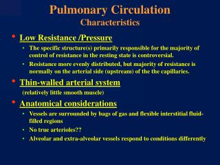
Pulmonary circulation What is the cardiovascular system? The heart is a double pump heartarteries arterioles veinsvenules capillaries
In mediastinum – behind sternum and pointing left, lying on the diaphragm It weighs 250-350 gm (about 1 pound) Heart’s position in thorax Feel your heart beat at apex 2
Pericardium(see next slide) Starting from the outside… 4 Without most of pericardial layers
Circulation Patterns Figure 21-18
Superiorvena cava 7 Capillaries of Head and arms Pulmonaryartery Pulmonaryartery Capillariesof right lung Capillariesof left lung Aorta 9 6 2 3 3 4 11 Pulmonaryvein Pulmonaryvein 5 LEFT ATRIUM 1 RIGHT ATRIUM LEFT VENTRICLE RIGHT VENTRICLE 10 Aorta Inferiorvena cava Capillaries ofabdominal organsand legs 8
Circulatory System Functions • Carry O2 to cells and CO2 away from cells • Deliver nutrients through body (after absorption in small intestine) • Carry liquid wastes away from cells (H2O, salt, urea) • Help in fighting infections • Temperature regulation
The Pulmonary Circuit Figure 21-19
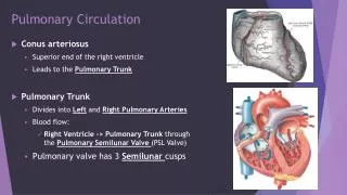
The Pulmonary Circuit (1 of 3) • Deoxygenated blood arrives at heart from systemic circuit: • passes through right atrium and ventricle • enters pulmonary trunk
The Pulmonary Circuit (2 of 3) • At the lungs: • CO2 is removed • O2 is added
The Pulmonary Circuit (3 of 3) • Oxygenated blood: • returns to the heart • is distributed to systemic circuit
Three circuits • Pulmonary • Blood goes from heart to lungs to pick up oxygen and release carbon dioxide • Systemic • Blood pumped out of heart to the rest of the body • Sound of heart (lub/dub) made by valves closing • Coronary • Heart muscle itself supplied with oxygen, nutrients, etc.
Requirements of gas exchange • Moist environment • O2 and CO2 must be dissolved to diffuse • Lungs, gills, moist surface (slime) help • Surface area--large area allows for more diffusion • Cleaned and filtered • Warmed
Movement of air into body • Nose--external opening to allow entry • Air is filtered, cleaned, warmed, moistened • Enters a series of tubes • Protected by cartilage to keep tubes firm/open • Mucus--traps foreign particles • Cilia-- “sweep” foreign material away from lungs to be swallowed (or spit/coughed)
Diffusion of gases • O2 concentration is higher in alveoli than blood: oxygen diffuses into blood • Remember High Conc. -> Low Conc. • At body cells O2 concentration is higher in blood: oxygen diffuses out of blood
Oxygen Transport • O2 diffuses from alveoli to the pulmonary capillaries. • O2-rich blood travels to heart and pumped to the body • O2 diffuses into cells. In tissues O2 levels are lower triggers Hb to release O2 • In tissues, CO2 makes blood more acidic and causes Hb to change shape. • CO2 diffuses from cells to blood. Travels to heart in form of Bicarbonate ions (HCO3-) • Heart pumps blood to lungs where CO2 is released in in gaseous form and then expelled.
LYMPH What is lymph ? Tissue fluid (interstitial fluid) that enters the lymphatic vessels
LYMPHATIC SYSTEM Essentially a drainage system accessory to venous system larger particles that escape into tissue fluid can only be removed via lymphatic system
Functions of the Lymphatic System • Reabsorbs excess interstitial fluid: • returns it to the venous circulation • maintain blood volume levels • prevent interstitial fluid levels from rising out of control. • Transport dietary lipids: • transported through lacteals • drain into larger lymphatic vessels • eventually into the bloodstream. • lymphocyte development, and the immune response.
Components of the Lymphatic System 24-33 • Lymph • Lymphatic Vessels • Lymphatic Capillaries • Lymphatic Vessels • Lymphatic Trunks • Lymphatic Ducts • Lymphatic Organs • Thymus • Lymph Nodes • Spleen • Tonsils • Lymphatic cells
Lymph Vessels Lymphatic capillaries – Lymphatic collecting vessels Lymphatic trunks – Lymphatic ducts –
Lymphatic Capillaries 24-36 Features of structure: Blind end Single layer of overlapping endothelial cells More permeable than that of blood capillary Absent from avascular structures, brain, spinal cord splenic pulp and bone marrow
Lymphatic Capillaries – Lacteals 24-37 The small intestine contains special types of lymphatic capillaries called lacteals. Lacteals pick up not only interstitial fluid, but also dietary lipids and lipid-soluble vitamins. The lymph of this area has a milky color due to the lipid and is also called chyle.
Lymphatic Vessels 238 Features of structure • Three layered wall but thinner than vein, • More numerous valves than in vein • Interposed by lymph nodes at intervals • Arranged in superficial and deep sets
LYMPH TRUNKS • right and left jugular trunks • right and left subclavian trunks • right and left bronchomediastinal trunks • right and left lumbar trunks • intestinal trunk
LYMPHATIC DUCTS 24-40 Right lymphatic duct • Formed by union of right jugular, subclavian, and bronchomediastinal trunks • Ends by entering the right venous angle
LYMPHATIC DUCTS • Thoracic duct • Begins in front of L1 as a dilated sac, the cisterna chyli, • formed by left and right lumbar trunks and intestinal trunk • Enter thoracic cavity & ascends • Travels upward, veering to the left at the level of T5
THORACIC DUCT….. At the root of the neck, it turns laterally arches forwards and descends to enter the left venous angle before termination, it receives the left jugular, Subclavian and broncho-mediastinal trunk
DRAINAGE PATTERN RIGHT LYMPHATIC DUCT -Receives lymph from right half of head, neck, thorax and right upper limb, right lung, right side of heart, right surface of liver • THORACIC DUCT - Drains lymph from lower limbs, pelvic cavity, abdominal cavity, left side of thorax, and left side of the head, neck and left upper limb
Lymphatic Cells 24-44 • Also called lymphoid cells. • Located in both the lymphatic system and the cardiovascular system. • Work together to elicit an immune response. • Types of lymphatic cells are: • macrophages • epithelial cells • dendritic cells • lymphocytes
LYMPHATIC ORGANS Primary organs • Red bone marrow • Thymus gland Secondary organs • Lymph nodes • Lymph nodules • Spleen
Lymph Nodes 24-46 Small, round or oval located along the pathways of lymph vessels. length from 1 - 25 millimeters Typically found in clusters receive lymph from many body regions. Lymph nodes are also found individually throughout the body tissues.
Lymph node Features • Bean-shaped bodies • With afferent vessels (entering at the periphery) and efferent lymph vessels(emerging at the hilus) • Arranged in groups, along the blood vessels or the flexural side of the joint • Divided into superficial and deep groups
Regional Lymph Node is the lymph node where the lymph of the organ or part of the body drainge to firstly Sentinel Lymph Node Regional Lymph drainage
Location Left epigastric region between 9th-11th rib in line of 10th rib Largest lymphatic organ in the body. Can vary considerably in size and weight Function Spleen
THYMUS Features • Consists of two elongated lobes • Is a large organ in the fetus • Occupies the thoracic cavity behind the sternum • Secrete lymphopoietin