Download
1 / 28
280 likes | 568 Vues
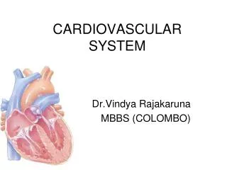
Cardiovascular System: Blood and Blood Vessels. Blood Vessels. The channels through which blood is distributed to body tissues This is a closed system Two types of vessels Pulmonary vessels – blood to and from the lungs Systemic vessels – blood to and from the tissues. Arteries.

E N D
Blood Vessels • The channels through which blood is distributed to body tissues • This is a closed system • Two types of vessels • Pulmonary vessels – blood to and from the lungs • Systemic vessels – blood to and from the tissues
Arteries • Carry blood AWAY from the heart • Pulmonary arteries – carry deoxygenated blood from right ventricle to the lungs • Systemic arteries – carry oxygenated blood from the left ventricle to the body tissues
Walls of arteries • Three layers called tunics • tunica externa– outermost • -attaches vessel to tissues • - elastic & collagen • b.tunica media – middle • -smooth muscle & elastic tissue • -thickest layer • tunica interna– innermost • -lines blood vessels • -continuous with the endocardium
Two types of arteries • 1. Elastic Arteries – largest arteries -thin walled • -many elastic fibers • –serve as pressure reservoirs to help pump blood when ventricles are relaxed • -Ex. Aorta, brachiocephalic, common carotids, subclavians
Muscular Arteries • -thick walled • –more smooth muscle than elastic fibers • –influence vasoconstriction (vessels getting smaller) & vasodilation (vessels getting bigger) to control blood flow • –Ex. Brachials & radials
Arterioles • Smallest arteries that deliver blood to capillaries • Help to regulate blood flow into tissue capillaries
Capillaries • Smallest & most numerous of blood vessels • found near almost every cell in the body • Extremely thin • Primary function is diffusion • Diffusion allows for the exchange of materials between the blood in the capillaries and the adjacent tissue cells (movement from regions of high concentration to low concentration) • Ex. – CO2, O2, glucose, hormones, amino acids, etc.
Capillary distribution - varies with metabolic activity of cells • Muscles, liver, kidneys – have a large capillary network due to their high metabolic activity (need lots of O2 and nutrients) • Tendons, ligaments – have few capillaries (not as active) • Cornea, lens of eye, cartilage – no capillaries
Bulk flow: filtration & reabsorption • Helps to maintain fluid balance and is based on pressure differences • Filtration – fluid movement from capillaries to cells • Reabsorption – fluid movement from cells to capillaries • Edema – swelling • -due to excessive filtration or inadequate reabsorption • - caused by high blood pressure or leaky capillaries
Venules • Collect blood from the capillaries and drain it into the veins
Veins • carry blood TOWARD the heart • Pulmonary veins – transport oxygenated blood from the lungs to the left atrium of the heart • Systemic veins – transport deoxygenated blood from the body tissues to the right atrium
Walls of veins also have three layers • Little smooth muscle & no elastic fibers • Larger diameter than arteries • Contain valves that project into the lumen toward the heart to prevent backflow • Serve as blood reservoirs – 60% of total blood in body is in veins • Allows blood to be quickly diverted to parts of the body that need it (vasoconstriction)
Blood flow velocity • Aorta (Fastest) • Capillaires (Slowest) • Vena Cavas (Faster) • Allows blood to spend more time in capillaries for diffusion • Normal circulation time for one drop of blood is one minute (to the aorta and back)
Blood pressure • Ventricular contraction puts pressure on walls of blood vessels • the greater the distance from the left ventricle, the lower the blood pressure (excludes elastic arteries) • Arteries have higher blood pressure than veins • Normal blood pressure is 120/80 mm Hg • Women usually have higher BP than men…8-20mmHg higher
Measured as systolic/diastolic • Systolic – pressure caused by ventricles contacting • Diastolic – pressure caused by ventricles relaxing • Sphygmomanometer – instrument used to measure BP • Stethoscope – 1st sound heard is the flow of blood through the collapsed artery (systolic); then you listen for no sound…this represents blood freely flowing through the artery (diastolic)
Blood pressure depends on: • Volume of Blood • - normal volume is ~ 5 L • -increase in volume– increase in BP • (water retention) • -decrease in volume-decrease in BP • (hemorrhaging) • 2.Vessel Resistance • a. Vessel radius • -if the radius is smaller, resistance is greater, BP increases (blockages, occlusions, cholesterol, blood clots, etc.)
b. Blood Viscosity • - thickness of blood • - the higher the viscosity, the higher the resistance, the higher the BP (dehydration) and vice versa • c. Vessel length • - the greater the length, the greater the resistance, the higher the BP
Regulation of blood pressure • Baroreceptors • - specialized cells that line arteries, veins and the right atriumand can sense changes in pressure • - 3 important ones: carotid sinus, aortic arch & right atrium • - If BP increases, baroreceptors stretch and trigger parasympathetic response which causes heart rate to decrease & BP to drop • -If BP decreases, barorecptors stretch less and trigger sympathetic response which causes heart rate to increase & BP to increase
Chemoreceptors • - measure changes in the chemical composition of blood • ex.; acidosis – an increase in H+ concentration (causes an increase in sympathetic impulses; increases heart and breathing rates) • hypoxia - lowered O2 availability • hypercapnia – excess CO2
Blood • a connective tissue • 3 functions • 1. transportation – O2, CO2, nutrients, waste, hormones • 2. regulation – body temp, pH, fluid balance • 3. protection – against disease & loss of blood (clotting)
Composition of blood • Centrifugation - process of separating blood into its components • Plasma – straw colored liquid portion of the blood (55%) • - 92% water, 8% solutes • - solutes are antibodies, enzymes, hormones, nutrients, electrolytes, waste products • 2. Formed elements–cells & cell fragments • -formed by a process called hemopoiesis
2. Erythrocytes – red blood cells • - tiny, biconcave discs; no nucleus • - 120 day life cycle • - function is to transport oxygen (does this by 4 O2 molecules binding to hemoglobin ) • - anemia results when there is a decreased amount of RBC’s or hemoglobin • erythropoiesis – formation of RBC’s • stimulated by the hormone erythropoietin
Leukocytes - white blood cells • - larger than RBC’s; have a nucleus • - active in immune response • Complete Blood Count (CBC) • - looks at all the different types of WBC’s present in the blood • -high WBC count – indicates ongoing infection • -low WBC count – indicates immune system failure
Different types of wbc’s • 1. neutrophils – engulf bacteria by phagocytosis (inflammation) • 2. monocytes– macrophages (TB) • 3. lymphocytes – control immune response • (T cells), kill cancer cells, attack transplanted tissue (cancer, HIV) • 4. eosinophils– attack antigen-antibody complexes (chicken pox) • 5. basophils– release histamine causing inflammatory response (allergies, allergic reaction
4. Thrombocytes – platelets • - cell fragments that function in clotting • Clotting of Blood • hemostasis – stop hemorrhaging • Vascular spasm – smooth muscle contraction • Platelet plug formation -platelets stick to damaged vessel -platelets change shape -chemicals released that cause other platelets to stick to the site (platelet plug) • Coagulation – blood clotting • - plug formation requires 13 proteins • - missing just 1 protein – blood cannot clot (hemophilia)
Blood type • -inherited characteristic • ABO antigens – RBC’s have antigens on their surface • can have A antigen, B antigen, both or neither • - your immune system recognizes the correct antigen and will attack foreign antigens (produces antibodies) • Rh antigens – either have it (Rh +) or you don’t (Rh-)
Blood types • Eight Blood Types • A+, A-, B, B-, AB+, AB-, O+, O- • Universal Recipient • Can receive any type of blood because it has all antigens on its surface • AB+ • Universal Donor • Can give blood to any other type because it has NO antigens on its surface • O-