Download
1 / 18
180 likes | 354 Vues
Analytical Techniques Utilizing Antibodies. flow cytometry gel electrophoresis immunoprecipitation (IP) immunoblotting microscopy immunofluorescence (IFA) electron microscopy ELISA. Immunofluorescence. Used to : localize antigens to specific cells or subcellular structures

E N D
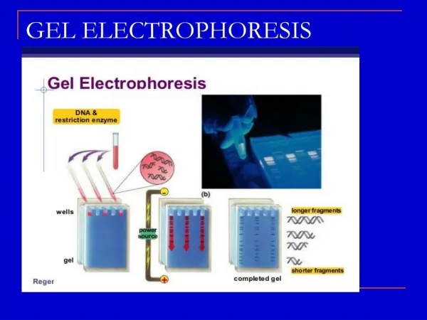
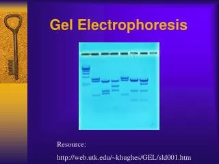
Analytical Techniques Utilizing Antibodies • flow cytometry • gel electrophoresis • immunoprecipitation (IP) • immunoblotting • microscopy • immunofluorescence (IFA) • electron microscopy • ELISA
Immunofluorescence • Used to: • localize antigens to specific cells or subcellular structures • detect and quantify antibody • General Procedure: • incubate cells or tissue with antibody • detect Ag-Ab complex with conjugated secondary antibody • fluorescence (examine under UV illumination) • enzyme (substrate forms precipitate)
Preparation of Cells • section and mount tissues • grow adherent cells on micro-scope slides or cover slips • affix suspension cells on microscope slides • carry out incubations in suspension • ± fixation • organic solvents • paraformaldehyde • <0.1% glutaraldehyde • ± permeabilization (eg., detergents) • surface labeling of unfixed cells IFA Protocol 1. Prepare cells or tissue. 2. Incubate 1o antibody. 3. Wash. 4. Incubate 2o antibody. 5. Wash. 6. View under UV illumination.
epifluorescence bright field • bright image against dark background • corresponds to location of antigen
Detergent Permeabilization + 0.1% TX-100
DAPI stains only DNA Counter Staining with Fluorescent Dyes EtBr stains DNA and RNA
Dual-Labeling Experiments • determine extent of co-localization • use 1o antibodies from different species and 2o antibodies labeled with different fluorochromes • label 1o antibodies with different fluorochromes
Immuno-Electron Microscopy • prepare samples • optimize fixation conditions • use resins that polymerize at RT • ‘float’ grids on drops (1o and 2o abs, washes) • surface of section accessible to antibodies (± 'etching') • 2o-Ab conjugated with colloidal gold • size ranges from 5-15 nm • enzyme linked (electron dense precipitate)
Ultrastructure vs. Labeling • fixation conditions preserving ultrastructure lead to loss of labeling • cryo-electron microscopy • special microtome and stage
Accessibility Problems • ultrasmall gold (<1 nm) • + silver enhancement • ±pre-embedding
IFA Characterizing Antibodies • Use same antigen with different antibodies • Quantify by serial dilutions
Conventional ELISA • bind antigen to 96-well microplate (or membrane) • neg. (and pos.) controls • purity? • incubate with 1o and 2o antibodies • use soluble chromogenic substrates in 96-well plates • quantify Ab or Ag
ELISA Variations • radio-immunosorbent assay (RIA)
Generic Immunoassay Procedure • form antibody-antigen complex • detect Ag-Ab complex • labeled anti-antibody (2o Ab) • labeled protein A or G • directly label 1o Ab • Fluorochromes • fluorescein • rhodamine • Enzyme Crosslinking • AP • HRP • Radiolabeling • iodination • metabolically (mAbs) • Biotinylation
Biotin-Avidin Detection Systems • label 1o- or 2o-Ab with biotin • detect with avidin labeled with marker • high affinity may increase sensitivity • more steps