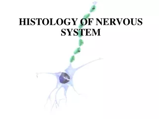
Unit IV NERVOUS SYSTEM HISTOLOGY
210 likes | 361 Vues
Biology 220 Anatomy & Physiology. Unit IV NERVOUS SYSTEM HISTOLOGY. Chapter 11 pp. 387-396. E. Gorski/ E. Lathrop-Davis/ S. Kabrhel. Functions. Sensory – recognize changes in environment [stimuli] Integration – analysis of sensory information, storage of information, decisions

Unit IV NERVOUS SYSTEM HISTOLOGY
E N D
Presentation Transcript
Biology 220 Anatomy & Physiology Unit IVNERVOUS SYSTEM HISTOLOGY Chapter 11 pp. 387-396 E. Gorski/ E. Lathrop-Davis/ S. Kabrhel
Functions • Sensory – recognize changes in environment [stimuli] • Integration – analysis of sensory information, storage of information, decisions • Motor – initiates impulses to effectors [muscles or glands] that do work)
Organization Fig. 11.2, p. 388
Cells Neurons and Supporting Cells • neurons • produce impulses to transfer information • amitotic (mostly), high metabolic rates, long-lived • supporting cells – support, protect, nurture neurons • neuroglia (glial cells) • astrocytes, oligodendrocytes, ependymocytes, microglia, satellite cells, Schwann cells (neurolemmocytes) • in CNS, tumors arise from abnormal divisions of glial cells
Supporting Cells in the CNS • Astrocytes – connect neurons to blood supply • Microglia – phagocytic Fig. 11.3, p. 389
Supporting Cells in the CNS • Oligodendrocytes – produce myelin sheath • Ependymal cells – epithelial lining of brain ventricles and central canal of spinal cord • produce cerebrospinal fluid Fig. 11.3, p. 389
Supporting Cells in PNS • Satellite cells –surround neuron cell bodies in ganglia • Schwann cells (neurolemmocytes) • form myelin sheaths around larger nerve fibers • play role in regeneration of nerve fibers Fig. 11.3, p. 389
Myelination (Myelin Sheath) • formed by oligodendrocytes (CNS), Schwann cells (PNS) • surrounds some axons (fibers) in CNS and PNS • composed of lipids and proteins (neurolemma = cell membrane of Schwann cell in PNS) • nodes of Ranvier = spaces between sheath cells • importance of myelin sheath: • increase speed of impulse conduction • decrease energy required (Na+/K+ pump only active at nodes) Multiple sclerosis– destruction of myelin sheath in CNS diminishes impulse conduction
Development of Myelin Sheath Fig. 11.5, p. 393
Neurons General: • most are amitotic (no cell division) • communicate with each other at synapses • neuron-neuron • neuroeffector junction (NEJ) • neuromuscular junction (NMJ) • neuroglandular junction (NGJ) • high rate of aerobic respiration
Neuron Cell body (perikaryon) • contains nucleus • rich in ribosomes & rough ER (Nissl bodies) • produces proteins for export to axon or dendrite • lots of mitochondria • neurofibrils
Neuron Processes Dendrites (d) • bring depolarization toward cell body • no myelin Axons (a) • generally take action potential (impulse) away form cell body • myelinated or unmyelinated • axon hillock • telodendria • synaptic end bulb d Fig. 11.4, p. a
Classification of Neurons • Based on structure – number of processes extending from cell body • unipolar • bipolar • multipolar • Based on function – type & direction of information • sensory • motor • association (interneurons)
Major Structural Classes Unipolar neurons • Unipolar neurons • dendrites short, lead to myelinated axon (central and peripheral processes) before cell body • generally sensory neurons within peripheral nervous system Table 11.1, p. 395
Major Structural Classes Bipolar Neurons • Bipolar neurons • one axon, one dendrite • sensory, including retina of eye and olfactory mucosa Table 11.1, p. 395
Major Structural Classes Multipolar Neurons • Multipolar : • one axon, several dendrites • interneurons, motor neurons • may be myelinated or unmyelinated Table 11.1, p. 395
Functional Classes of Neurons Based on type and direction of information (impulse) transmission • Sensory: • afferent (brings sensory info to CNS) • most unipolar or bipolar • Interneurons: • integration between sensory & motor in CNS • most multipolar • Motor: • efferent (goes toward/to effector) • most multipolar
Other Definitions • Nerve fiber:long axon (primarily in PNS) • Nerve:bundle of neuron processes (fibers) in PNS • Tract:bundle of neuron processes in CNS • Ganglion (ganglia): cluster of cell bodies in PNS • Nucleus (nuclei): cluster of cell bodies in CNS • White matter: myelinated nerve processes in CNS • outside in spinal cord; inside in brain • Gray matter: unmyelinated nerve processes in CNS • inside in spinal cord, outside in brain
Nerve Structure • Nerves = bundles of neuron processes (axons) in PNS • Coverings: • endoneurium: wraps individual fibers (over myelin sheath); composed of areolar CT • perineurium: groups fibers into bundles called fascicles; composed of dense irregular CT • epineurium: encloses fascicles, arteries, veins; composed of dense irregular connective tissue Fig. 13.2, p. 481
Peripheral Nerve Types • Sensorynerves carry afferent fibers only • Motornerves carry efferent fibers only • Mixednerves carry both kinds of fibers 1 = epineurium 2 = perineurium 3 = endoneurium