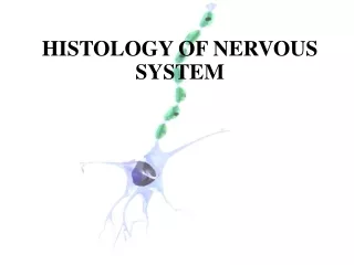
Central Nervous System Histology Slide Orientation
320 likes | 924 Vues
Central Nervous System Histology Slide Orientation. Spinal cord. Dorsal horns: Interneurons Ventral horns: Motor neurons Lateral horns: Sympathetic neurons Parasympathetic (S2-4) neurons DRG: Sensory (pseudounipolar) neurons

Central Nervous System Histology Slide Orientation
E N D
Presentation Transcript
Central Nervous System Histology Slide Orientation
Spinal cord Dorsal horns: Interneurons Ventral horns: Motor neurons Lateral horns: Sympathetic neurons Parasympathetic (S2-4) neurons DRG:Sensory (pseudounipolar) neurons Autonomic ganglia: Post ganglionic neurons with unmyelinated axons. Sympathetic: paravertebral ganglia Parasympathetic: In organs
65-2 Lumbar Spinal Cord, H&E Dura mater D.R.G. Spinal Cord Myelinated nerves
#65-1N Spinal cord and DRG Dorsal horn Lateral horn Ventral horn Dorsal root ganglion (DRG)
66a Thoracic Spinal Cord Dorsal median sulcus Dorsal nucleus of Clarke (proprioceptive) Dorsal horn Lateral horn Ventral horn Ventral median fissure
66a Thoracic Spinal Cord Neurons in Lateral Horn Neurons in ventral Horn
65-1N Motor neurons, glial cells and neuropil Glial cells Neuropil
#74 Sympathetic ganglion cells #75 Parasympathetic ganglion cells Seminal vesicle
Sensory and Autonomic Neurons(DRG) (Parasympathetic)
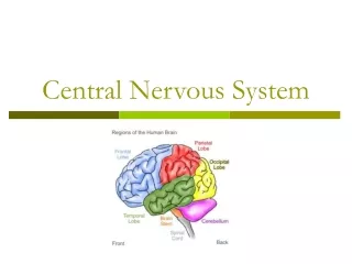
Brain Cerebrum Sulcus (sulci) Gyrus (gyri) 3rd ventricle Cerebellum 4th ventricle Pons Medulla oblongata
Cerebrum and Cerebellum 76b 77
#77 Cerebellum #76b Cerebrum C: Cortex of gray matter W: White matter C C C W W W
#77 Cerebellum Cortex White matter
#77 Cerebellum ML GL WM Purkinje neurons (cells)
#76 Cerebrum White matter Cortex of gray matter
#76 Cerebral cortex Ross and Pawlina, p.311
CerebellumCerebrum C: Cortex of gray matter W: White matter Purkinje cells (neurons) C C Pyramidal cells (neurons) W W
Hippocampal Formation Posterior View Lateral View
LIMBIC LOBE (Cortical Areas) Cortex Type Cingulate gyrus & Isthmus Neocortex (six layers) Subcallosal area Parahippocampal Gyrus Paleocortex (3-5 layers) Uncus Hippocampus Archicortex (3 layers) (Deep within parahippocampal gyrus)
Hippocampal Formation Dentate GyrusHippocampusSubiculum Martin: Neuroanatomy Figs. 15-3, 4, 14
#NP004N Hippocampal regionCA: Cornus ammonis Choroid plexus CA2 Fornix Lateral ventricle CA3 Dentate gyrus Hippocampus CA1 Subiculum
3 layers of Dentate gyrusArchicortex (3 layers), as opposed to Neocortex (6 layers) Molecular layer Molecular layer (dendrites of granule cells) Granule cell layer (neurons) Polymorphic layer Polymorphic layer (interneurons)
3 layers of Hippocampus(Archicortex) Polymorphic layer Pyramidal cell layer Molecular layer Pyramidal cells