Plasmid DNA Isolation
300 likes | 481 Vues
Plasmid DNA Isolation. Exercise 8. Experiment Goals. Extraction of plasmid DNA from E. Coli Analyze plasmid DNA by agarose gel electrophoresis and spectrophotometer. Plasmid. Plasmids.

Plasmid DNA Isolation
E N D
Presentation Transcript
Plasmid DNA Isolation Exercise 8
Experiment Goals • Extraction of plasmid DNA from E. Coli • Analyze plasmid DNA by agarose gel electrophoresis and spectrophotometer
Plasmid Plasmids • A plasmid is an extrachromosomal DNA molecule separate from the chromosomal DNA which is capable of replicating independently of the nuclear DNA. E. coli Chromosome • Circular and double-stranded • Plasmid size varies from 1 to over 100 kbp • The number of identical plasmids within a single cell can be zero, one, or even hundreds under some circumstances.
Classification of plasmids by function There are five main classes • Fertility-F-plasmids, Facilitate bacterial conjugation • Resistance-(R)plasmids, which contain genes that can build a resistance against antibiotics or poisons. • Col-plasmids, which contain genes that code for bacteriocins, proteins that can kill other bacteria. • Degradative plasmids, which enable the digestion of unusual substances, e.g., toluene or salicylic acid. • Virulence plasmids, which turn the bacterium into a pathogen.
Plasmid Applications • The plasmids used in transformation typically have three important elements: • A cloning site (a place to insert foreign DNAs) • An origin of replication • A selectable marker gene (e.g. resistance to • ampicillin)
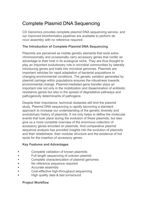
Plasmid DNA isolation • Plasmid DNA isolation requires separation of this DNA from the chromosomal DNA in the bacterial cell as well as from the polysaccharides, lipids and proteins that constitute the cell.
Methods for Plasmid Isolation • There are several methods to isolate plasmid DNA from bacteria: • Miniprep • Can be used to quickly find out whether the plasmid is correct in any of several bacterial clones. The yield is a small amount of impure plasmid DNA, which is sufficient for analysis by restriction digest and for some cloning techniques. • Maxiprep/bulkprep • Much larger volumes of bacterial suspension are grown from which a maxi-prep can be performed. Essentially this is a scaled-up miniprep followed by additional purification. This results in relatively large amounts (several micrograms) of very pure plasmid DNA.
Overnight Culture Suspension • Pick a single colony and inoculate in 5 ml of LB containing 20 mg/l ampicilin • Incubate overnight at 37oC • Centrifuge 1.5 ml of broth containing cells in a tube • Discard supernatant
Plasmid DNA isolation • Inactivation of Bacteria • Lysis of cells/ denaturation of DNA • Precipitation of DNA • Separate plasmid DNA from contaminants • Precipitation of Plasmid DNA • Precipitation of proteins • Precipitate Plasmid DNA
1- Inactivation of Bacteria • Resuspend cell pellet in 100 µl of GTE buffer (50mM Glucose, 25 mM Tris-Cl & 10mM EDTA, pH 8) • Glucose is added to increase the osmotic pressure outside the cells • Tris is a buffering agent • EDTA protects the DNA from degradative enzymes • Vortex gently if necessary
2- Lysis of cells/ denaturation of DNA Add 200 µl of NaOH/ SDS lysis solution, invert tube 6-8 times 1. Sodium dodecyl sulfate • Dissolves membranes • Binds to and denatures proteins 2. NaOH • NaOHrupture the cell and also denatures the DNA into single strands
3- Precipitation of DNA • Immediately add 150 µl of 5 M potassium acetate solution (pH 4.8) 1. Potassium acetate / acetic acid solution • Neutralizes NaOH (renatures plasmid DNA) • Converts soluble SDS to insoluble PDS sodium dodecyl sulfate (SDS) potassium dodecyl sulfate (PDS) • Precipitate the genomic DNA • Centrifuge for 1 minute at high speed
4- Separate plasmid DNA from contaminants Separate plasmid DNA from contaminants by centrifugation • Supernatant contains: - Plasmid DNA - Some cellular constituents • Sediment contains: - PDS - Lipids - Proteins - Chromosomal DNA
5- Precipitation of Plasmid DNA • Transfer supernatant layer to a clean tube and add 0.5 ml of isopropanol on ice for 10 minutes • Centrifuge at top speed for 1 minute Add 0.5 ml of isopropanol to supernatant Supernatant Centrifuge Incubate for 10 min. on ice Pellet • Remove supernatant, dissolve pellet in 0.4 ml TE buffer • Add 10 µl of RNAse solution, vortex & incubate at 37oC for 20 – 30 min.
Mix thoroughly with an equal volume of organic solvent Aqueous Centrifuge phenol, chloroform, Organic 6- Precipitation of proteins • Add 300 µl of phenol/ chloroform/ Isoamyl alcohol • Vortex vigorously for 30 seconds • Centrifuge at full speed for 5 minutes
Supernatant Absolute ethanol & ammonium acetate precipitated DNA Centrifuge Pellet 7- Precipitate Plasmid DNA • Remove supernatant to a clean tube • Add 100 µl of 7.5 M ammonium acetate & 1 ml of absolute ethanol to precipitate the plasmid DNA, incubate on ice • Mix and then centrifuge at full speed for 5 minutes •Wash pellet with 75% Ethanol (to remove salts), & dry pellet • Dissolve pellet with TE (or other aqueous solution)
Quantifying Plasmid DNA • Quantify DNA using UV absorbance • DNA UV absorbance peaks at 260 nm • protein UV absorbance peaks at 280 nm • The ratio of the absorbance at 260 nm/280 nm is a measure of the purity of a DNA sample from protein contamination; it should be between 1.7 and 2.0 • The ratio of the absorbance at 260 nm/230 nm is a measure of the purity of a DNA sample from organics and/or salts; it should be about 2.0. Low 260/230 ratio indicates contamination by organics and/or salts
Assessing plasmid preparation Verify by restriction digestion Run undigested plasmid to see if it is mostly supercoiled denatured supercoiled
Analyzing Plasmids • After digestion, the restriction fragments are separated by gel electrophoresis. • The banding pattern from the restriction fragments provide a genetic “fingerprint” of the plasmid and gene insert.
Lane 1: Supercoiled (lower band) and open circular form (upper band) • Lane 2: Multimeric forms of supercoiled plasmid DNA Lane 3: Linearized form of a plasmid after restriction digestion with EcoRI. Lane 4: Sample contaminated with bacterial chromosomal DNALane 5: EcoRI digestion of a sample contaminated with bacterial genomic DNA which gives a smear above the plasmid DNA.