Human Genomic DNA Isolation
130 likes | 303 Vues
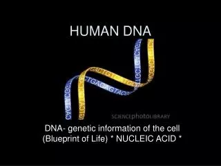
Human Genomic DNA Isolation. Zelha Nil Nov 2009. DNA Structure . Composed of nucleotides : A, T, G, C Synthesized in 5’ to 3’ direction through formation of phosphodiester bonds betw deoxyribose & phosphate : Sugar - Phosphate backbone

Human Genomic DNA Isolation
E N D
Presentation Transcript
Human Genomic DNA Isolation Zelha Nil Nov 2009
DNA Structure • Composed of nucleotides: A, T, G, C • Synthesized in 5’ to 3’ directionthroughformation of phosphodiesterbondsbetwdeoxyribose & phosphate: Sugar-Phosphatebackbone • Doublehelix: H-bondingbetwcomplementarybases • Specificsequence of bases: protein structure & geneticinheritance
Organization of Human Genome • Human genome: Total genetic information (DNA content) in human cells • Nuclear genome: 99.9995% of the total genetic information • Mitochondrial genome: the remaining 0.0005% • Nuclear genome • Chains of DNA organized into chromosomes • Human: 3x10⁹ bp packed into 23 chromosomes, 2n=46 • Human chromosomes: 50-250 Mb in length • Each chromosome: Single long molecule of DNA
Homologous chromosomes One from each parent: non-identical Replication Sister chromatids Identical
Euchromatin: Lightly packed form of chromatin that is rich in gene concentration • Heterochromatin: Tightly packed form of DNA
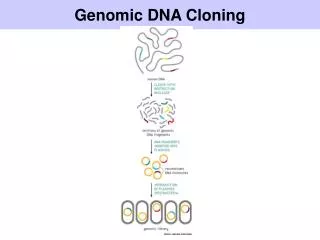
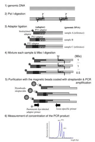
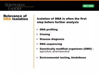
Why we isolate genomic DNA? • PCR; presence of sequencesoramplification of targetsequences (mutationalanalysis, cloning) • Southernblot; presence of sequences (DNA fingerprinting, cloning) • Sequenceanalysis (mutationalanalysis, sequencingthegenome) • DNA fragmentation; indication of apoptosis • RE digestion; RFLP (DNA polymorphisms), DNA fingerprinting, cloning, PCR
DNA polymorphisms • Definition: Differences in nucleotide sequence among individuals of a species • Result from • Point mutations • Random indels • Variable repeat numbers in a repetitive locus • Used for DNA fingerprinting • Repetitive DNA sequences • Restriction fragment length polymorhisms (RFLP)
RFLP analysis • Based on variability in restriction enzyme (RE) cut sites betw individuals • Single base changes in DNA • Introduce or delete a RE cutsite • For ex: A mutation changing the sequence AGATCC to GGATCC introduce a BamH1 site into that segment of DNA • Alterationin RE cut site: • Variation in the length of the fragments • Difference in the position of certain gel bands betw individuals
Common procedures • Phenol-chloroform extraction (manual) • Salting out (our protocol) • ASSIGNMENT: What are the differences betw these 2 methods & which one is more efficient or advantageous? What can be other methods alternative to these? (1 page)
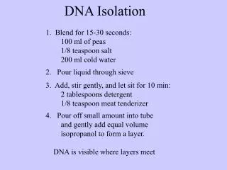
DNA isolation by salting out method • Put 750 µl blood, 750 µl TKM buffer and 10 µl Triton X-100 into 2 ml eppendorf tube, mix well by inversions. • Centrifuge at 1000g for 10 min at RT. • Discard supernatant slowly, save the pellet. • Add 750 µl TKM buffer and resuspend the pellet. • Centrifuge at 1000g for 10 min at RT. • Repeat the steps 3-5 2 more times. • Resuspend the pellet in 200 µl TKM buffer. • Add 20 µl of 10%SDS, mix well. • Incubate the samples at 58oC for 10 min.
Cont’d • Add 75 µl cold saturated NaCl. • Centrifuge at 14000g for 10 min at 4oC. • Save the supernatant (300 µl) into a new 1.5 ml eppendorf tube. • Add 2x volume (600 µl) of absolute ethanol, invert slowly several times. DNA is visible in this step. • Incubate the samples at -20oC for 30 min. • Centrifuge at 10000g for 10 min at 4oC. • Pour off the ethanol, let the eppendorfs dry under hood. • Add 200 µl TE buffer pH:8.0, resuspend. • Incubate at 37oC for at least 2 hours.
TKM buffer (TrisHCl, EDTA) Hypotonicbufferforlysis of RBC and WBC enhancedbyinversions. • Triton X 100, SDS (10% w/v) Detergentstosolubilizelipidsandproteins • SaturatedNaCl Nucleilysisbuffer • Absoluteethanol (96-100% v/v) Added 2-3 volumes of thesolutiontocollect DNA (orprecipitatenucleicacids) fromaquousphases since nucleicacidstendtoinsolubilize in ethanol. • TE buffer (10mM Tris, 1mM EDTA, pH 7.5) Dissolve DNA andstoragebuffer