Download
1 / 38
400 likes | 850 Vues
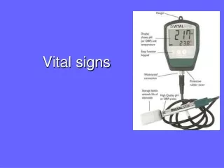
Vital signs. Outline. Vital Signs Definition Temperature Pulse Rate Respiratory Rate Blood Pressure Pain. Vital sign. physical signs that provide data to determine a person’s state of health

E N D
Outline • Vital Signs Definition • Temperature • Pulse Rate • Respiratory Rate • Blood Pressure • Pain
Vital sign • physical signs that provide data to determine a person’s state of health • indicate an individual is alive, such as temperature, pulse rate, respiratory rate (TPR), and blood pressure (BP).
Measuring Body Temperature • Purposes • 1-To establish baseline data for subsequent evaluation . • 2-To identify whether the core body temperature is within normal range . • 3-To determine changes in the core body temperature in response to specific therapies ( antipyretic medication , immunosuppressive drugs, invasive procedure ) • 4-To monitor clients at risk for imbalanced body temperature ( clients at risk for infection , or diagnosis of infection , or those who have been exposed to temperature extreme)
Types of Thermometers Electronic thermometers • Provide readings in less than 60 seconds • most accurate if placed in sublingual pocket • There is a sensor on the end of the thermometer that touches the body part and reads the body’s temperature.
Types of Thermometers Tympanic membrane thermometer • measures the temperature inside of the ear. • It will read the infrared heat that comes from inside of the ear. • Especially appropriate for infants and young children • Readings are obtained in 2 seconds or less
Types of Thermometers Glass and mercury thermometers • a glass tube with mercury inside of the tube. • The tube goes underneath the tongue and the body temperature will cause the mercury to rise inside the tube. • DO NOT just throw away a mercury thermometer.
Assessment : • 1-Clinical signs of fever . • 2-Clinical signs of hypothermia • 3-Site most appropriate for measurement . • 4-Factors that may alter body temperature.
Planning Preparation of equipment : 1-Thermometer 2-Thermometer cover . 3-Water- soluble lubricant for a rectal temperature . 4-Disposable gloves . 5- Towel for axillary temperature . 6-Tissue /wipes
Implementation Preparation: Check that all equipments functioning well . Performance : 1- Introduce self , verify the client’s identity , explain to the client what will you do, why and how ? 2- Hand washing . 3-Provide for client’s privacy . 4-Position the patient according to the method will be practiced ( lateral or sim’s position for rectal temperature ) 5-Place the thermometer as the following :
Evaluation Compare the temperature measurement to baseline data , normal range of age of the client and the client’s previous temperature . Analyze considering time of day and any additional influence factors and other vital signs .
Assessment of peripheral Pulse • Purpose : • To establish baseline data for subsequent evaluation. • To identify whether the pulse rate is within normal range . • To determine whether the pulse rhythm is regular and the pulse volume is appropriate . • To determine the equality of corresponding peripheral pulse on each side of the body . • To monitor and assess changes in the client’s health status . • To monitor client’s at risk for pulse alteration ( heart disease , cardiac arrhythmia . • To evaluate perfusion to the extremities
Assessment `1-Clinical signs of cardiovascular alterations as: (dyspnea, cyanosis, palpitations , syncope , cool skin ) 2- Factors that may alter pulse rate ( e.g. emotional status , physical activity ) . 3- Which site is most appropriate for assessment based on a purpose .
Assessment of apical pulse : Position the patient in comfortable supine position or in a sitting position . Locate the apex of heart
Planning • Equipment : • -Watch with a second hand or indicator. • Implementation • Performance : • 1- Introduce self , verify the client’s identity , explain to the client what will you do, why and how ? • 2- Hand washing . • 3-Provide for client’s privacy . 4- Select the pulse point . Normally , the radial pulse is taken unless it can’t be exposed . • 5- Position the patient in a rest position
Implementation : 6- Palpate and count the pulse . Place 3 or 2 middle fingers lightly and squarely over the pulse point . 7- Count for 15 seconds and multiply by 4 . 8- Record the pulse on the worksheet . 9- Assess the pulse rhythm and strength . 10- Document the pulse rate on the patient’s record . 11- Hand wash
Evaluation 1-Compare the pulse rate to baseline data or normal range for age of the client . 2- Relate pulse volume , rate to other vital signs , pulse rhythm and volume to other baseline data and health status . 3- Conduct appropriate follow up such as notifying the primary care giver or giving medication .
C-Assessment of Respiration : Purposes : To acquire baseline data against which future measurements can be compared . To monitor abnormal respiration and respiratory patterns and identify changes . To monitor respirations before or following the administration of general anesthetic or any medication that can influences respiration . To monitor clients at risk for respiratory alterations .
Assessment : Skin and mucous membrane color ( cyanosis or pallor ) Positions assumed for breathing ( using of orthopneic position). Signs of cerebral anoxia ( irritability , restlessness drowsiness or loss of consciousness ) . Chest movement . Activity tolerance. Chest pain . Dyspnea Medication that affect respiration .
Planning Equipment: Watch with a second or indicator . Implementation : Preparation: For a routine assessment of respiration , determine the client’s activity schedule and choose a suitable time to monitor the respirations . A client who has been exercising will need to rest for a few minutes to permit the accelerated respiratory rate to return to normal .
Implementation : 1- Introduce self , verify the client’s identity , never to notify the patient that you will assess respiration 2- Hand washing . 3-Provide for client’s privacy . 4-Observe and count the respiratory rate . 5- Observe the respiration for depth by watching the movement of the chest , observe for regularity . 6- Document the respiratory rate , rhythm and depth in an appropriate record
Evaluation Relate respiratory rate to other vital signs , in particular pulse , relate respiratory rhythm ,and depth to baseline data and health status . Report to the primary care provider a respiratory rate significantly above or below the normal range and any notable change in respiration from a previous assessment . Conduct appropriate follow up such as administering oxygen, or other medications
Assessment of Blood Pressure Purpose : 1-To obtain a baseline measure of arterial blood pressure for subsequent evaluation . 2- To determine the client’s hemodynamic status . 3- To identify and monitor changes in blood pressure resulting from a disease processes .
Equipment Sphygmomanometer Aneroid Mercurial Stethoscope
Sphygmomanometer Pediatric Adult
Parts of stethoscope • Earpieces- should fit snugly and follow the natural curve of the ear canal, point toward the face when it is in place • Tubing- 12-18 inches long, longer tubing decreases the transmission of sound waves
Parts of a stethoscope • Diaphragm= circular, flat surface- transmits high pitched sounds ( Bowel, lung, heart sounds • Bell= bowl shaped- transmits low pitched sounds (heart and vascular sounds)
Assessment 1- Signs & symptoms of hypertension ( headache , ringing in the ears , flushing of the face ,nosebleeds, fatigue ). 2- Signs & symptoms of hypotension ( tachycardia , dizziness, mental confusion , restlessness cool and clammy skin, pale or cyanosis ) 3- Factors affecting blood pressure ( stress , activity , pain and time of last caffeine .) 4- Some blood pressure cuffs contains latex . Assess the client for latex allergy and obtain a latex –free cuff if indicated .
Planning Equipment : 1- stethoscope 2-Blood pressure cuff (appropriate size) Sphygmomanometer Preparation : 1-Ensure that the equipment is intact and functioning well 2- Make sure that the client has not smoked within 30 minutes
Implementation Preparation : 1-Ensure that the equipment is intact and functioning well 2- Make sure that the client has not smoked within 30 minutes Performance : 1- Introduce self , verify the client’s identity , explain to the client what will you do, why and how 2- Hand washing . 3-Provide for client’s privacy .
4-Take the accurate reading of blood pressure and Document the finding in the client’s record . 5-Hand wash
Evaluation 1- Relate blood pressure to other vital signs , to baseline data . 2- Report any significant changes in client’s blood pressure . 3- Conduct appropriate follow up , medication administration .