Download
1 / 49
500 likes | 708 Vues
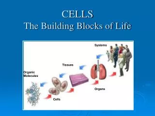
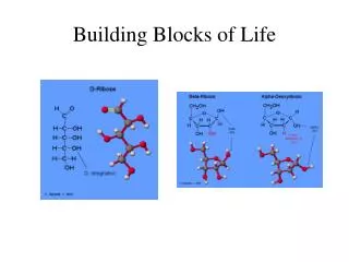
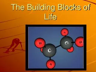
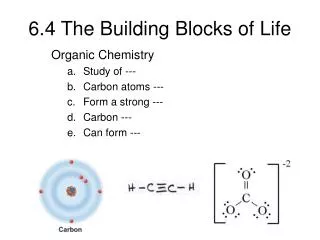
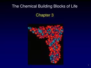
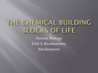
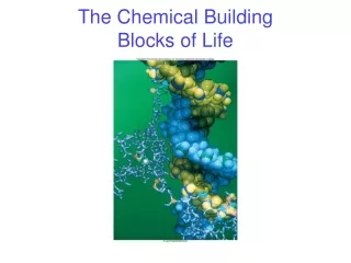
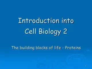
Introduction into Cell Biology 2 The building blocks of life - Proteins. Intro into cell biology 2. Molecular Organisation of a cell. Fig. 1.7. Building Blocks of Life -> Different Shapes. Proteins Amino Acids are linked by peptide bonds.

E N D
Introduction into Cell Biology 2The building blocks of life - Proteins
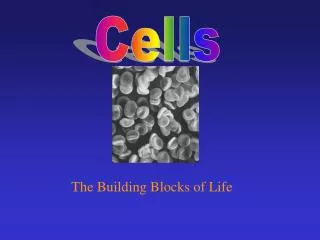
Intro into cell biology 2 Molecular Organisation of a cell
Proteins Amino Acids are linked by peptide bonds
20 Natural Occurring Amino Acids are divided into groups according to their side chains:
Cys can cross-link between 2 polypeptide chains -> Disulfide bridge
Proteins are Polypeptides Direction of a Protein
Interactions between side chains and backbone -> Fold of a protein (3D structure)
Noncovalent interactions within and between biological molecules
Secondary Structure: 1. α – Helix 2. β-Strands -> β-Sheets 3. Loops and Turns
α-helical coiled coil proteins: Form superhelix Found in myosin, tropomyosin (muscle), fibrin (blood clots), keratin (hair) Examples of α-Helical Proteins: Hair
Examples of α-Helical Proteins: Muscle α-helical coiled coil proteins: Form superhelix Found in myosin, tropomyosin (muscle), fibrin (blood clots), keratin (hair)
Examples of β-sheet Proteins: Fatty acid binding protein -> β barrels structure OmpX: E. coli porin Antibodies
Turns and Loops: Loops in Receptors Turn
Quaternary Structure: Polypeptide chains assemble into multisubunit structures Cell-surface receptor CD4 Tetramer of hemoglobin
Protein Folding Folding is a highly cooperative process (all or none) Folding by stabilization of Intermediates
Misfolded protein -> Alzheimer Protein fibrillation
Function of Proteins Specific binding of ligands -> Immunoglobins
Function of Proteins Conformational change of lactoferrin upon binding of Fe Conformational change induced by Calcium
Function of Proteins Activation by modification GFP fluorescent: Rearrangement and oxidation of Ser-Tyr-Gly
Function of Proteins Model of enzymatic reaction mechanism
Proteins Key properties: • Proteins are linear polymersbuilt of Amino Acids • Proteins contain many functional groups (i.e.. side chain of AA) • Proteins interact with proteins and with other biological molecules to form complexes • Proteins can bind and/or modify other molecules • Proteins can be rigid or can have regions with high flexibility
Enzyme Kinetics • Enzymes DO NOT shift the equilibria but enhance the rates of the reactions (lower the activiation energy!!!)
X‡ intermediate H Reaction coordinate Transition state • Unstable state of maximum energy • Not an intermediate • Metastable state • Intermediates are species that appear in a reaction mechanism but not in the overall balanced equation. DH‡ H DH0 Reaction coordinate
rate = D[A] D[B] rate = - Dt Dt Reaction Kinetics Thermodynamics – does a reaction take place? Kinetics – how fast does a reaction proceed? Reaction rate is the change in the concentration of a reactant or a product with time (M/s). A B D[A] = change in concentration of A over time period Dt D[B] = change in concentration of B over time period Dt Because [A] decreases with time, D[A] is negative. 13.1
A B time rate = D[A] D[B] rate = - Dt Dt 13.1
Suppose an enzyme were to react with a substrate, giving a product. Basic problem of enzyme kinetics S + E P + E If we simply applied the law of mass action to this reaction, the rate of reaction would be a linearly increasing function of [S]. As [S] gets very big, so would the reaction rate. This doesn’t happen. In reality, the reaction rate saturates.
Michaelis and Menten In 1913, Michaelis and Menten proposed the following mechanism for a saturating reaction rate k1 k2 S + E ES P + E k-1 Complex. product
Michaelis-Menten Kinetics • When [S] << KM, the reaction increases linearly with [S]; I.e. vo = (Vmax / KM ) [S] • Very little [ES] is formed • When [S] = KM, vo = Vmax /2 (half maximal velocity); this is a definition of KM: the concentration of substrate which gives ½ of Vmax. This means that low values of KM imply the enzyme achieves maximal catalytic efficiency at low [S]. • When [S] >> Km, vo = Vmax Where activity measurements should be performed: 1. [S] very high 2. all enzyme bound in [ES] complex
Michaelis-Menten Kinetics When the enzyme is saturated with substrate, the reaction is progressing at its maximal velocity, Vmax. Combing the steady-state assumption (d[ES]/dt=0) with the conservation condition ([E]T=[E] + [ES]) vo leads to the Michaelis-Menten Equation of enzyme kinetics: where Km is KM= (k-1 + k2)/k1
Michaelis-Menten Kinetics What is Vmax and KM ? • KM gives an idea of the range of [S] at which a reaction will occur. The larger the KM, the WEAKER the binding affinity of enzyme for substrate. • Vmax gives an idea of how fast the reaction can occur under ideal circumstances.
Michaelis-Menten Kinetics Determination of Enzyme kinetics -> Measure activity (velocity) at different substrate concentrations Determine activity of an Enzyme -> Measure at substrate concentration of above 10KM -> no substrate limitation [E]T=[ES]
Michaelis-Menten Kinetics How to measure activity of an enzyme using photometrical method ? Lambert Beer law: A= c ε l Where A is the absorbance c is the concentration (mol/L) ε is the molar absorption coefficient (L/mol cm) l is the path length of sample (cm) Rate = activity = Δc/Δt -> ΔA/Δt = l εΔc/Δt activity = rate (unit) -> Δc/Δt = (ΔA/Δt)/lε Definition: 1 Unit of an enzyme will catalyse the reaction of 1 μmol of substrate within 1 min at certian pH and Temperature. Measure ΔA/Δt (change in absorption/min) for a special enzyme and a high substrate concentration -> from that you can calculate activity of that enzyme at special Temp., solvent, pH, pressure.
Michaelis-Menten Kinetics How do determine experimentally KM and Vmax ? (y= d + k x) Lineweaver-Burk plot Eadie-Hofstee plot