Download
1 / 53
560 likes | 622 Vues
Learn about the intricacies of protein degradation within cells and its impact on cellular processes and degenerative disorders. Explore the ubiquitin/proteasome pathway, autophagy, and the role of various proteins in regulating protein turnover.

E N D
Intracellular Protein Degradation Chris Weihl MD/PhD weihlc@neuro.wustl.edu Department of Neurology
Protein Degradation in the Cell Ub Nucleus Autophagy Ub Aggresome Ub UPS Ub Endocytosis
Consequence of impaired protein degradation • Protein aggregates • Ubiquitinated inclusions • Vacuolation • Damaged organelles • Secondary impairment in other cellular processes • Cell Death • Underlying pathogenesis of degenerative disorders (neurodegeneration, muscle degeneration, liver degeneration, lung disease, aging)
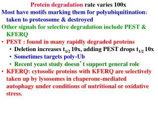
Protein Degradation • Turnover of protein is NOT constant • Half lives of proteins vary from minutes to infinity • “Normal” proteins – 100-200 hrs • Short-lived proteins • regulatory proteins • enzymes that catalyze committed steps • transcription factors • Long-lived proteins • Special cases (structural proteins, crystallins)
May depend on tissue distribution • Example: Lactic Acid Dehydrogenase • Tissue Half-life • Heart 1.6 days • Muscle 31 days • Liver 16 days • Protein degradation is a regulated process • Example: Acetyl CoA carboxylase • Nutritional state Half-life • Fed 48 hours • Fasted 18 hours Protein Degradation
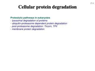
Protein Degradation • Ubiquitin/Proteasome Pathway • 80-90% • Most intracellular proteins • Lysosomal processes • 10-20% • Extracellular proteins • Cell organelles • Some intracellular proteins
UBIQUITIN • Small peptide that is a “TAG” • 76 amino acids • C-terminal glycine - isopeptide bond with the e-amino group of lysine residues on the substrate • Attached as monoubiquitin or polyubiquitin chains G K
Ubiquitination of proteins is a FOUR-step process • First, Ubiquitin is activated by forming a link to “enzyme 1”(E1). AMP • Then, ubiquitin is transferred to one of several types of “enzyme 2”(E2). • Then, “enzyme 3”(E3) catalizes the transfer of ubiquitin from E2 to a Lys e-amino group of the “condemned” protein. • Lastly, molecules of Ubiquitin are commonly conjugated to the protein to be degraded by E3s & E4s
The UPS is enormous! The UPS is enormous! The genes of the UPS constitutes ~5% of the genome • E1’s- 1-2 activating enzymes • E2’s- 10-20 conjugating enzymes • E3’s- 500-800 ubiquitin ligase- drives specificity • DUBs- 100 ubiquitin specific proteases- regulators of pathway The genes of the UPS constitutes ~5% of the genome • E1’s- 1-2 activating enzymes • E2’s- 10-20 conjugating enzymes • E3’s- 500-800 ubiquitin ligase- drives specificity • DUBs- 100 ubiquitin specific proteases- regulators of pathway
PROTEASOME COMPONENTS 20S Proteasome 19S Particle ATP 26S Proteasome
Hydrolysis peptide bonds after: hydrophobic a.a. =CHYMOTRYPSIN-LIKE - 5 acidic a.a. = (-) CASPASE-LIKE -1 basic a.a. = (+) TRYPSIN-LIKE -2
DEUBIQUITINATION De-ubiquitinating
Proteasome inhibitors proteasome ub-ub-ub-ub ub-ub-ub-ub ub-ub-ub-ub ub-ub-ub-ub proteasome ub-ub-ub-ub
Proteasome inhibition increases Usp14 ubiquitin-hydrolase activity Usp14 Uch37 Borodovsky, A et al EMBO J. 20:5187-96 2001
The proteasomal DUB Usp14 impairs protein degradation Lee, BH et al Nature 467:179-84 2010
Decrease steady-state levels of aggregate prone proteins in the absence of Usp14 Lee, BH et al Nature 467:179-84 2010
Lyosomal degradation • Autophagy
Autophagy • Lysosomal degradation of proteins and organelles • Occurs via three routes • Macroautophagy • Microautophagy (direct uptake of cellular debris via the lysosome) • Chaperone mediated autophagy (selective import of substrates via Hsc70 and Lamp2a)
Yeast Genetics meets Human Genetics • Identification of >50 autophagy essential proteins with mammalian homologs
Macroautophagy FOXO3 Beclin ATG7 Lysosome mTOR ATG5-ATG12-ATG16L1 Autophagosome Induction Nucleation Trafficking & Cargo loading Autolysosome Phagophore Sequestration Degradation Fusion “Autophagic Flux”
Genetic knockout of autophagy initiating proteins Complete loss of ATG5 leads to lethality
Tissue specific knockout of autophagy • Degeneration of CNS tissue; Hara et al 2006 • Hepatomegaly in Liver; Komatsu et al 2005 • Atrophy and weakness of skeletal muscle; Masiero et al 2009 • Pathologic similarities • Ubiquitinated inclusions • Aberrant mitochondria • Oxidatively damaged protein
Basal Autophagy • Autophagy has a “housekeeping” role in the maintenance of cellular homeostasis • Autophagy is responsible for the clearance of ubiquitinated proteins
Selective Autophagy • Aggregaphagy– p62/SQSTM1, Nbr1 • Mitophagy – Parkin, Nix • Reticulophagy – endoplasmic reticulum • Ribophagy – translating ribosomes • Xenophagy – e.g. Salmonella via optineurin • Lipophagy – autophagy mediated lipolysis • Performed by an expanding group of ubiquitin adaptors
p62 as an autophagic tool • p62 associates with ubiquitinated proteins and LC3 • p62 is an autophagic substrate
LC3 as an autophagic tool LC3-I (18kD) LC3-II (16kD) GFP-LC3 starved
Autophagosome proteins are elevated in IBMPFD 2 p62 protein levels (A.U) 1 0 Con WT RH9 RH12 2 LC3II protein levels (A.U) 1 0 Con WT RH9 RH12 Ju et al, JCB 2009
Autophagosome accumulate in IBMPFD cells Ju et al, JCB 2009
Why do autophagosomes accumulate? • Upregulation of functional autophagosomes • Decrease in autophagosome degradation or “autophagic flux” • Phagophore closure • Autophagosome-lysosome fusion • Absence of functional lysosomes
IBMPFD mutant VCP impairs “autophagic flux” Ju et al, JCB 2009
IBMPFD has “blocked” autophagy Ub Nucleus
Rapamycin as an inducer of autophagy • Immunosuppressant used to treat transplant rejection • Inhibits the mTOR pathway • mTOR integrates extrinsic growth signals and cellular nutrient status and energy state • Active mTOR • Protein synthesis and cell growth • Inactive mTOR (or rapamycin treatment) • Inhibition of protein synthesis and increased autophagic degradation of protein