Download
1 / 30
300 likes | 326 Vues
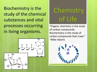
The relationship of cellular chemistry (biochemistry) to cellular structure.

E N D
The relationship of cellular chemistry (biochemistry) to cellular structure.
There are several important themes that transcends the chemistry and bring the importance of understanding the cell biological differences between eukaryotes and prokaryotes. These themes are all part of the evolution of eukaryotes. The evolution of internal membrane structures gives rise to the organelles referred to as the cytomembranes,while the another organelle group belongs to the endosymbionts.How they arose and how the endosymbionts evolved has changed greatly since Lyn Margulis original thesis.What is the advantages of compartmentation?
The genes for most cellular proteins are located in the nuclear genome. The mRNA escapes the nucleus to be translated on ribosomes. What happens next?
Questions from previous slide; • How are proteins targeted to the right organelle? • Can the same protein be found in the more than one compartment? • How then are they located in both spaces? Are they analogous or homologous?
The cell membrane What are the functions of the cell membrane (PM)? Why is it a PM? Compare and contrast the prokaryotic and eukaryotic PM? What is derived (evolved) from the a proto-bacterial PM?
The extracellular matrix (ECM) is the extracellular part of multicellular structure that typically provides structural and biochemical support to the surrounding cells Because multicellularity evolved independently in different multicellular lineages, the composition of ECM varies between multicellular structures; however, cell adhesion, cell-to-cell communication and differentiation are common functions of the ECM. The animal extracellular matrix includes the interstitial matrix and the basement membrane. Interstitial matrix is present between various animal cells (i.e., in the intercellular spaces). Gels of polysaccharides and fibrous proteins fill the interstitial space and act as a compression buffer against the stress placed on the ECM. Basement membranes are sheet-like depositions of ECM on which various epithelial cells rest.
Mammalian organisms are complex associations of cells into tissues and organs. How are cells in tissues held together? How do they communicate with one another? The proteins on these cells PM, are then associated with specific faces of the tissue cell, How do they find their location?
Prof. C. DeDuve, M.D., Biochemist, Nobel Prize Recipient Physiology & Medicine 1974
Fractionation of cellular organelles by Differential cfg. What is the advantage of Density Gradient cfg as opposed to Differential Cfg. Prof DeDuve discovered both the method of density grad cfg, marker enzymes, the lysosome and the hydrogenosome. He described the relationship between the ER, Golgi and lysosome.
Potential mechanisms of signal orientation in the translocon. (A) Retention of the more positive flanking sequence of the signal by interaction with negative charges on the cytosolic side at or near the translocon. (B) Loop insertion of the polypeptide followed by inversion of reverse signal–anchors with a more negative N-terminus. (C) Head-on insertion with subsequent inversion of cleavable signals and type II signal–anchors with a more positive N-terminal flanking sequence. For simplicity, the SRP receptor has been omitted. The signal sequence is schematically drawn as a helix; yet, its conformation is not known during targeting and reorientation.
The entrance of a protein into the mitochondria presents unique problems for transport. There is an inner and outer membrane, most proteins are associated with the inner membrane or transmembraneous proteins of the inner membrane.
This is a view for how proteins that are translocated into the ER, then is transported to the Golgi. The vesicular Traffic is bidirectional.
Peroxisomes (also called microbodies) are organelles found in virtually all eukaryotic cells. They are involved in the catabolism of very long chain fatty acids, branched chain fatty acids, D-amino acids, polyamines, and biosynthesis of plasmalogens, i.e. ether phospholipids critical for the normal function of mammalian brains and lungs.They also contain approximately 10% of the total activity of two enzymes in the pentose phosphate pathway, which is important for energy metabolism. It is vigorously debated if peroxisomes are involved in isoprenoid and cholesterol synthesis in animals. Other known peroxisomal functions include the glyoxylate cycle in germinating seeds ("glyoxysomes), glycolysis in trypanosomes ("glycosomes"), and methanol and/or amine oxidation and assimilation in some yeasts. Peroxisomes can be derived from the endoplasmic reticulum and replicate by fission. Peroxisome matrix proteins are translated in the cytoplasm prior to import. Specific amino acid sequences (PTS or peroxisomal targeting signal) at the C-terminus (PTS1) or N-terminus (PTS2) of peroxisomal matrix proteins signals them to be imported into the organelle.
TCR CD28 CD4 CD3 PiP2 InsP3 + DAG PI3-K Raf-1 Ras fyn PLC p110 Lck p85 MKK 2 1 Lck [Ca2+] PKC ZAP-70 3 Grb-2 p75 SOS Vav Calcium signaling PKC signaling Grb-2 I-kβ C3G Shc p116 NF-kβ Crk1 Cbl Ras/MAPK signaling JNK calcineurin ERK-1.2 NFATc NFAT AP-1 NF-kβ NFAT Fos/Jun (AP-1) Differential cytokine genes transactivated
Molecules are taken up from outside the cell in endocytotic vesicles, which fuse with early endosomes. Membrane components are recycled as the early endosomes mature into late endosomes. Transport vesicles carrying acid hydrolases from the Golgi apparatus then fuse with late endosomes, which mature into lysosomes as they acquire a full complement of lysosomal enzymes. The acid hydrolases dissociate from the mannose-6-phosphate receptor when the transport vesicles fuse with late endosomes, and the receptors are recycled to the Golgi apparatus.
The process of Secretion has two senarios, 1. constitutive were there is constant secretion of the vesicle content ie albumin by the liver or 2. regulated secretion, for instance of insulin by the b-cells of the pancreas. Also, there are cargo in vesicles being transported From Golgi to late endosomes to join with the lysosome.
Profs. Otto Warburg, M.D./Ph.D. & Sir Hans Krebs M.D., Biochemists, Nobel Recipients
Although differing in detail, both of these studies Karlberg O, et al The dual origin of the yeast mitochondrial proteome. Yeast. 2000. Marcotte EM,et al . Localizing proteins in the cell from their phylogenetic profiles. PNAS 2000, come to similar general conclusions about the origin of the yeast mitochondrial proteome. In particular, the two studies - which both consist fundamentally of similarity searches - identify three categories of yeast mitochondrial proteins: 'prokaryote-specific' (50-60% of the total), 'eukaryote-specific' (20-30%) and 'organism-specific', or 'unique' (about 20%). Prokaryote-specific mitochondrial proteins are defined as those that have counterparts in prokaryotic genomes; eukaryote-specific mitochondrial proteins have counterparts in other eukaryotic genomes but not in prokaryotic genomes; and organism-specific mitochondrial proteins are ones so far unique to S. cerevisiae. In addition, both studies point out that this classification correlates with the known or inferred functions of the proteins in each category: prokaryote-specific mitochondrial proteins predominantly perform roles in biosynthesis, bioenergetics and protein synthesis, whereas eukaryote-specific mitochondrial proteins function mainly as membrane components and in regulation and transport.
Where do all of these pathways arise from? • Why are most of the genes for these pathways in the nucleus? • If many of these genes are not α-proto-bacterial then where did they arise form? • Was the mitochondria free living or parasitic?
Cartoon of the hydrogenosomal metabolism of Nyctotherus ovalis. The hydrogenosomes of Nyctotherus ovalis perform a chimericmetabolism that exhibits mitochondrial and hydrogenosomal traits. The basis is provided by a ciliate mitochondrial metabolism. However, the TCA cycle is incomplete, and it is likely that the TCA cycle is used in the reductive way in the hydrogenosomes of Nyctotherus ovalis. The function of complex I has been substantiated by the demonstration of a proton gradient that is sensitive against several inhibitors of a mitochondrial complex I and insensitive against cyanide and antimycin, which interfere with the pumping of mitochondrial complex III/IV. The excretion of succinate (besides acetate, lactate and ethanol) strongly suggests a function of mitochondrial complex II as fumarate reductase, which is likely to accept electrons from complex I through a rhodoquinone 9 (Tielens et al. 2002; Boxma et al. 2005). The Fe-hydrogenase and the “fumarate respiration” regulate the organellar NADH pool allowing reoxidation to NAD. The links to the fatty acid and amino acid metabolism are substantiated by the identiWcation of a number of key genes. Thepresence of an active ATP synthase remains to be established byfurther experiments. (1) Pyruvate dehydrogenase. (2) Malatedehydrogenase. (3) -Ketoglutarate dehydrogenase. (4). Succinyl CoA synthase. (5) Succinate dehydrogenase/fumarate reductase.(6) Phosphoenolpyruvate carboxykinase (PEPCK). (7) 2-Oxoglutarate/malate translocator. (8) Acetyl/propionyl CoA carboxylase( α-subunit)
Separation of functional complexes of the respiratory chain. The outer mitochondrial membrane is first removed by treatment with the detergent digitonin. Fragments of inner membrane are then obtained by osmotic rupture of the mitochondria, and the fragments are gently dissolved in a second detergent. The resulting mixture of inner membrane proteins is resolved by ion-exchange chromatography into different complexes (I through IV) of the respiratory chain, each with its unique protein composition, and the enzyme ATP synthase (sometimes called Complex V). The isolated Complexes I through IV catalyze transfers between donors (NADH and succinate), intermediate carriers (Q and cytochrome c), and O2, as shown. In vitro, isolated ATP synthase has only ATP-hydrolyzing (ATPase), not ATP-synthesizing, activity.
Topology of the NE. Inner and outer nuclear membranes (INM and ONM, respectively) are separated by the ER lumen or perinuclear space (PNS). The nuclear lamina interacts with NE proteins and chromatin. INM proteins link the NE to chromatin and the lamina. ONM proteins provide a connection from the nucleus to the cytoskeleton. The lamin B receptor (LBR) interacts both with B-type lamins and chromatin-associated heterochromatin protein 1 (HP1) in conjunction with core histones. Members of the LEM (lamina-associated protein 2 [LAP2], emerin, MAN1)-domain family (pink) bind to lamins and interact with chromatin through barrier-to-autointegration factor (BAF). SUN proteins (SUN 1 and 2) interact with nesprins in the ONM, thereby forming so-called LINC complexes that establish connections to actin and intermediate filaments in the cytoplasm. Nurim is a multi-pass membrane protein with unknown function. Proteomic approaches have identified ~60 putative transmembrane proteins (NETs), most of which remain uncharacterized. .
Proteins are transported through the nuclear pore complex in two steps. In the example shown, a protein with a classical basic amino acid-rich nuclear localization sequence (NLS) is recognized by importin α, which forms a complex with importin β. Importin β binds to the cytoplasmic filaments of the nuclear pore complex, bringing the target protein to the nuclear pore. The protein and importin α are then translocated through the nuclear pore complex in a second, energy-requiring step, which requires GTP hydrolysis by the Ran protein.