Download
1 / 12
120 likes | 322 Vues
Alteration of Anterior Chamber Angle after Implantation of Iris-fixated Phakic Intraocular Lens. Takefumi Yamaguchi, Kazuno Negishi, Megumi Saiki, Kenya Yuki, Nanae Kawaguchi, Kazuo Tsubota Keio University, School of Medicine. Introduction.

E N D
Alteration of Anterior Chamber Angle after Implantation of Iris-fixated Phakic Intraocular Lens Takefumi Yamaguchi, Kazuno Negishi, Megumi Saiki, Kenya Yuki, Nanae Kawaguchi, Kazuo Tsubota Keio University, School of Medicine
Introduction The iris-fixated PIOL implantation has been proved to be a effective and predictable refractive technique for treatment of high myopia. However, severe postoperative complications have been reported. One of the concerns is that PIOLs inevitably cause some postoperative intraocular structure change; pupil ovalization, anterior chamber depth, and crystalline lens rise. We evaluated the ACA after PIOL implantation.
Objective We evaluated the alteration of anterior chamber angle (ACA) after implantation of iris-fixated intraocular lens (PIOL) for myopia and its postoperative effects on anterior chamber structure and inflammation. Subjects Twenty eyes of 11 patients (7 female and 4 males) The mean age; 38.4±11.1years old (26~58) The mean preoperative spherical equivalent ; –9.8±4.5 D (-18.25~-2.25D) (Artisan Model 204 were implanted for myopia in all eyes.)
Methods The Scheimpflug image was obtained before and 1 month after implantation of PIOL using the Pentacam (Oculus, Germany). The angle at 2, 3, 4, 8, 9, 10 o’clock were measured by a protractor. Data Analysis ・ Alteration of ACA (ΔACA) ・ Postoperative intraocular pressure (IOP) at 1 month ・ Effects of ΔACA on change in anterior chamber depth (ACD) and anterior chamber volume (ACV) ・ Postoperative flare at 1 month and 1 year measured by laser flarimetry (FC-2000, Kowa, Tokyo, Japan)
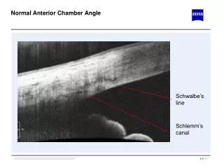
Example of the Scheimpflug image after implantation of iris-fixated PIOL A B The anterior chamber angle is narrowed near the iris-fixation point (A) compared with the other points (B).
Pre- and postoperative ACA degree
Alteration of ACA N=20 ΔACA=(preoperative ACA) – (postoperative ACA) ACA: Anterior chamber angle
Pre- and postoperative IOP IOP There was no significant difference between before and after surgery. (p>0.05)
Effects of ΔACA on ACV and ACD ΔACD mm3 ΔACV mm3 P=0.08 P=0.74 degree degree ΔACA ΔACA ΔACV=(Preoperative ACV) – (Postoperative ACV) ACA: Anterior chamber angle ACV:Anterior chamber volume ACD:Anterior chamber depth ΔACD=(Preoperative ACD) – (Postoperative ACD)
Postoperative flare and ΔACA 1 month after surgery 1 year after surgery (photons/mm2) (photons/mm2) p=0.0071 p=0.004 degree Correlation between |ΔACA| and aqueous flare A, 1 month and B, 1 year after surgery. (Pearson’s correlation coefficient A, r=0.573, p=0.0071 1 month B, r=0.700, p=0.004 1 year after surgery.) |ΔACA|=| (average preoperative ACA) – (average postoperative ACA) | (degree) ACA: anterior chamber angle
Discussion Partial narrowing of the angle near the fixation points was induced by implantation of the iris-fixated PIOL and that postoperative acute and chronic anterior chamber inflammation had a significant correlation with the amount of the alteration of the angle. The angle alteration was thought to be caused by pinching the iris by the claw of the PIOL. However the amount of the angle alteration varied between individuals. The difference between patients could be due to surgical factors such as the amount of pinched iris and the position of the PIOL, and the individual patient’s factors such as iris hysteresis (stiffness of the iris). Further evaluation of the iris change is necessary for the iris-fixated PIOL and other types of phakic IOLs.
Conclusion Partial narrowing of the ACA was induced after implantation of PIOL for high myopia, which is correlated with postoperative acute and chronic anterior chamber inflammation. It would be desirable to observe ACA over long-term, especially in patients with large angle alterations.