Download
1 / 51
510 likes | 610 Vues
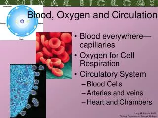
The heart and circulation Transportation- oxygen and carbon dioxide red blood cells Regulation-hormones, temperature Protection- against blood loss (clotting); infection (immune system). Components of the circulatory system Cardiovascular system- heart and blood vessels

E N D
The heart and circulation Transportation- oxygen and carbon dioxide red blood cells Regulation-hormones, temperature Protection- against blood loss (clotting); infection (immune system)
Components of the circulatory system Cardiovascular system- heart and blood vessels Lymphatic system- lymph nodes, lymphatic vessels
Electrical activity of the heart Myocardial cells beat automatically Action potential is usually originated in sinoatrial node Spontaneous depolarization (pacemaker potential) diffusion of calcium through slow channels threshold- fast calcium channels open, voltage regulated sodium channels open repolarization produced through diffusion of potassium
Electrocardiogram Conduction of electrical potentials through heart P wave- atrial depolarization QRS- ventricular depolarization beginning of systole T wave- repolarization of the ventricles beginning of diastole
Regulation of cardiac rate Rhythm is set by the SA node Sympathetic nerves epinephrine and norepinephrine stimulate opening of calcium and sodium channels; increase cardiac rate Parasympathetic (vagus) nerves acetylcholine promotes opening of potassium channels; reduces cardiac rate
Heart muscle cannot sustain contraction Long refractory periods- heart cannot be stimulated until it has relaxed from previous contraction Arrhythmias- something affects the cardiac cycle; treatment depends on what it is Fast Na channel blockers Slow Ca channel blockers -adrenergic receptor blockers
Arrhythmias Bradycardia- slow rate (less than 60 bpm) Tachycardia- fast rate (more than 100 bpm) Can occur normally; is abnormal if rate increases during rest (ectopic pacemakers) Flutters- extremely rapid contractions Fibrillation- different groups of fibers are activated so coordinated pumping of chambers is not possible
Blood vessels- arteries and veins Arteries, arterioles, capillaries Veins and venules Arteries are more muscular Veins have valves
Veins Veins can expand to accommodate increasing amounts of blood; arteries can’t Venous pressure is low compared to arterial pressure Blood flow through veins is facilitated by: contraction of skeletal muscles valves that prevent backflow
Atherosclerosis Damage to endothelium “Fatty streaks” (macrophages and lymphocytes) Fibrous plaques High blood cholesterol, LDL contribute to atherosclerosis HDL also help transport cholesterol, but do not contribute to atherosclerosis
Lymphatic system Fluid transport from tissues Fat transport from intestines Immune response
Regulation of cardiac activity Cardiac output Blood flow Blood pressure
Cardiac output= stroke volume X cardiac rate (ml/min) (ml/beat) (beats/min) At 70 beats/min and 80 ml/beat, this comes to about 5.5 liters per minute— Equivalent to the total blood volume
Stroke volume regulated by End-diastolic volume amount of blood in ventricles before they begin to contract increases stroke volume Total peripheral resistance to blood flow in the arteries the higher the resistance, higher pressure heart compensates by beating more strongly Contraction strength of ventricle; proportional to stroke volume
Exercise reduces vagus inhibition and increases sympathetic nerve activity Cardiac control center in medulla oblongata coordinates this activity This in turn is regulated by higher brain activity and pressure (baroreceptors) in aorta and carotid arteries
Venous return At rest, most of the blood is in the veins veins can “give” more and hold more blood than arteries; venous pressure is much lower (2 mm Hg vs. 90-100 mm Hg mean arterial pressure) Venous pressure determines rate of blood return to the heart
Blood volume Extracellular fluid distributed between blood plasma and interstitial fluid Affected by: forces acting at capillaries (to draw fluid out of or into them) overall balance of water loss and gain
Regulation of blood volume by kidneys Filtration of blood- almost all of filtrate is reabsorbed by the kidneys (out of daily production of ca. 180L of filtrate, only about 1.5 L actually excreted) Various hormones acting on, or produced by, the kidneys (to be discussed later, but think about: what causes fluid to be retained or lost?)
Resistance to blood flow Related to pressure difference between the ends of the vessel Inversely related to resistance of blood flow through vessel In body, vasodilation in one organ system might be offset by vasoconstriction in another
Regulation of blood flow Sympathetic nervous system overall, increase in cardiac output and in peripheral resistance vasoconstriction in arterioles of viscera and skin vasodilation in skeletal muscles (depends on receptors) Parasympathetic- vasodilation effect confined to GI, genitalia, salivary glands
Paracrine regulation, e.g., inflammation localized vasoconstriction, dilation Intrinsic (autoregulation) myogenic- response to changes in blood pressure (constrict to protect blood vessels, dilate to improve blood flow) metabolic-oxygen, carbon dioxide levels local vasodilation
Regulation of blood flow to the heart Alpha and beta adrenergic receptors (constriction and dilation; norepinephrine and epinephrine) Also intrinsic regulation increased metabolic rate- oxygen need, accumulation of carbon dioxide, etc. smooth muscle stimulated to cause relaxation and dilation
How are aerobic requirements of heart met? Lots of capillaries Myoglobin releases oxygen during systole (blood flow is reduced at that time) capacity for aerobic respiration: extra mitochondria, enzymes Blockages in blood supply are corrected by angioplasty, bypass, etc.
Total increased blood flow more to muscles, less to skin and viscera Overall flow to brain is about the same Mainly due to increased cardiac rate With conditioning, stroke volume also increases
Blood flow to brain Intrinsic mechanisms maintain constant flow myogenic responses to changes in blood pressure sensitive to CO2 levels in arterial blood metabolic responses- local vasodilation Blood flow to skin is highly sensitive to action of sympathetic nervous system temperature sensitive
Blood pressure Blood flow resistance highest in arterioles Flow rate lowest in capillaries Blood pressure can be raised by: vasoconstriction of arterioles increase in cardiac output (higher cardiac rate or stroke volume) Various factors can affect these: kidneys, sympathetic nervous system, etc.
Pressure receptors (baroreceptors) Action potentials will increase or decrease as pressure rises or falls Baroreceptor reflex activated when blood pressure rises or falls. Activated when a person changes position Vasomotor control centers- constriction/dilation Cardiac control centers- cardiac rate
Blood pressure also regulated by: Atrial stretch receptors ADH release Renin-angiotensin-aldosterone ANF
Measurement of blood pressure sphygmomanometer Systolic/diastolic pressure, e.g., 120/80 exercise tends to raise systolic more changing position tends to affect diastolic Pulse pressure: systolic- diastolic reflects stroke volume drops in dehydration or blood loss Pulse rate reflects cardiac rate Mean arterial pressure= diastolic + 1/3 pulse pressure (indicator of peripheral resistance)
Pathophysiology of cardiovascular system Hypertension Secondary- results from known diseases (table 14.10) processes that affect blood flow; damage to tissue that results in release of vasoactive chemicals; damage to sympa- thetic nervous system, etc. Essential- accounts for most cases of hypertension
Increased total peripheral resistance Low renin secretion? High salt intake? Stress? Inability of kidneys to regulation salt and water excretion?
Consequences of high blood pressure Can damage cerebral blood vessels and lead to stroke Causes heart to work harder (harder to eject blood if peripheral resistance is high) Contributes to atherosclerosis Treatments are many and varied diet, diuretics, various receptor blockers
Shock due to loss of blood flow hypovolemic- blood LOSS septic- blood-borne infection; nitric oxide formation might be the culprit (vasodilator) anaphylactic- severe allergic reaction (histamine) cardiogenic- infarction causes extensive damage to heart muscle
Congestive heart failure- cardiac output is inadequate causes: heart disease, hypertension, electrolyte imbalance Digitalis increases contractility of heart muscle Diuretics lower blood volume Nitroglycerin is a vasodilator Make heart work more efficiently; reduce stress on heart