Bleeding Disorders
Bleeding Disorders. Morey A. Blinder, M.D. Associate Professor of Medicine and Pathology & Immunology. Objectives. Coagulation factor disorders and treatment Disorders of platelets and platelet transfusion Adjunctive drug therapy for bleeding.

Bleeding Disorders
E N D
Presentation Transcript
Bleeding Disorders Morey A. Blinder, M.D. Associate Professor of Medicine and Pathology & Immunology
Objectives • Coagulation factor disorders and treatment • Disorders of platelets and platelet transfusion • Adjunctive drug therapy for bleeding
Inherited bleeding disorders Hemophilia A and B vonWillebrands disease Other factor deficiencies Acquired bleeding disorders Liver disease Vitamin K deficiency/warfarin overdose DIC Coagulation factor disorders
Ecchymoses (typical of coagulation factor disorders)
Hemophilia A and B Hemophilia A Hemophilia B Coagulation factor deficiency Factor VIII Factor IX Inheritance X-linked X-linked recessive recessive Incidence 1/10,000 males 1/50,000 males Severity Related to factor level <1% - Severe - spontaneous bleeding 1-5% - Moderate - bleeding with mild injury 5-25% - Mild - bleeding with surgery or trauma Complications Soft tissue bleeding
Hemophilia Clinical manifestations (hemophilia A & B indistinguishable) Hemarthrosis (most common) Fixed joints Soft tissue hematomas (e.g., muscle) Muscle atrophy Shortened tendons Other sites of bleeding Urinary tract CNS, neck (may be life-threatening) Prolonged bleeding after surgery or dental extractions
Treatment of hemophilia A • Intermediate purity plasma products • Virucidally treated • May contain von Willebrand factor • High purity (monoclonal) plasma products • Virucidally treated • No functional von Willebrand factor • Recombinant factor VIII • Virus free/No apparent risk • No functional von Willebrand factor
Dosing guidelines for hemophilia A • Mild bleeding • Target: 30% dosing q8-12h; 1-2 days (15U/kg) • Hemarthrosis, oropharyngeal or dental, epistaxis, hematuria • Major bleeding • Target: 80-100% q8-12h; 7-14 days (50U/kg) • CNS trauma, hemorrhage, lumbar puncture • Surgery • Retroperitoneal hemorrhage • GI bleeding • Adjunctive therapy • amino caproic acid (Amicar) or DDAVP (for mild disease only)
Complications of therapy • Formation of inhibitors (antibodies) • 10-15% of severe hemophilia A patients • 1-2% of severe hemophilia B patients • Viral infections • Hepatitis B Human parvovirus • Hepatitis C Hepatitis A • HIV Other
Treatment of hemophilia B • Agent • High purity factor IX • Recombinant human factor IX • Dose • Initial dose: 100U/kg • Subsequent: 50 U/kg every 24 hours
von Willebrand DiseaseClinical features • von Willebrand factor Carrier of factor VIII Anchors platelets to subendothelium Bridge between platelets • Inheritance Autosomal dominant • Incidence 1/10,000 • Clinical features Mucocutaneous bleeding
vonWillebrand type Assay 1 2 3 vWF antigen ß Normal ßß vWF activity ßßßß Multimer analysis Normal Normal Absent Laboratory evaluation of von Willebrand disease Classification • Type 1 Partial quantitative deficiency • Type 2 Qualitative deficiency • Type 3 Total quantitative deficiency • Diagnostic tests:
Treatment of von Willebrand diseaseVaries by Classification • Cryoprecipitate • Source of fibrinogen, factor VIII and VWF • Only plasma fraction that consistently contains VWF multimers • Correction of bleeding time is variable • DDAVP (Deamino-8-arginine vasopressin) • Increases plasma VWF levels by stimulating secretion from endothelium • Duration of response is variable • Used for type 1 disease • Dosage 0.3 µg/kg q 12 hr IV • Factor VIII concentrate (Humate-P) • Virally inactivated product • Used for type 2 and 3
Vitamin K deficiency • Source of vitamin K Green vegetables Synthesized by intestinal flora • Required for synthesis Factors II, VII, IX ,X Protein C and S • Causes of deficiency Malnutrition Biliary obstruction Malabsorption Antibiotic therapy • Treatment Vitamin K Fresh frozen plasma
Vitamin K deficiency due to warfarin overdoseManaging high INR values Clinical situation Guidelines INR therapeutic-5 Lower or omit next dose; Resume therapy when INR is therapeutic INR 5-9; no bleeding Lower or omit next dose; Resume therapy when INR is therapeutic Omit dose and give vitamin K (1-2.5mg po) Rapid reversal: vitamin K 2-4 mg po (repeat) INR >9; no bleeding Omit dose; vitamin K 3-5 mg po; repeat as necessary Resume therapy at lower dose when INR therapeutic Chest 2001:119;22-38s (supplement)
Vitamin K deficiency due to warfarin overdoseManaging high INR values in bleeding patients Clinical situation Guidelines INR > 20; serious bleeding Omit warfarin Any life-threatening bleeding Vitamin K 10 mg slow IV infusion FFP ± factor rhVIIa (depending on urgency) Repeat vitamin K injections every 12 hrs as needed
Disseminated Intravascular Coagulation (DIC)Mechanism Systemic activation of coagulation Depletion of platelets and coagulation factors Intravascular deposition of fibrin Thrombosis of small and midsize vessels with organ failure Bleeding
Sepsis Trauma Head injury Fat embolism Malignancy Obstetrical complications Amniotic fluid embolism Abruptio placentae Vascular disorders Reaction to toxin (e.g. snake venom, drugs) Immunologic disorders Severe allergic reaction Transplant rejection Common clinical conditionsassociated with DIC
DICTreatment approaches • Treatment of underlying disorder • Anticoagulation with heparin • Platelet transfusion • Fresh frozen plasma
Liver Disease Decreased synthesis of II, VII, IX, X, XI, and fibrinogen Prolongation of PT, aPTT and Thrombin Time Often complicated by Gastritis, esophageal varices, DIC Treatment Fresh-frozen plasma infusion (immediate but temporary effect) Vitamin K (usually ineffective)
Coagulation cascade Intrinsic system (surface contact) Extrinsic system (tissue damage) XII XIIa Tissue factor XIa XI IX IXa VIIa VII VIII VIIIa X Xa V Va (Thrombin) IIa IIa II Fibrinogen Fibrin Vitamin K dependant factors
Laboratory Evaluation of the Coagulation Pathways Partial thromboplastin time (PTT) Prothrombin time (PT) Surface activating agent (Ellagic acid, kaolin) Phospholipid Calcium Thromboplastin Tissue factor Phospholipid Calcium Intrinsic pathway Extrinsic pathway Thrombin time Common pathway Thrombin Fibrin clot
Problems with blue-top tube Partial fill tubes Vacuum leak and citrate evaporation Problems with phlebotomy Heparin contamination Wrong label Slow fill Underfill Vigorous shaking Pre-analytic errors • Biological effects • Hct ≥55 or ≤15 • Lipemia, hyperbilirubinemia, hemolysis • Laboratory errors • Delay in testing • Prolonged incubation at 37°C • Freeze/thaw deterioration
Initial Evaluation of a Bleeding Patient - 1 Normal PT Normal PTT Abnormal Urea solubility Factor XIII deficiency Normal Consider evaluating for: Mild factor deficiency Monoclonal gammopathy Abnormal fibrinolysis Platelet disorder (a2 anti-plasmin def) Vascular disorder Elevated FDPs
Initial Evaluation of a Bleeding Patient - 2 Normal PT Abnormal PTT 50:50 mix is abnormal Repeat with 50:50 mix Test for inhibitor activity: Specific factors: VIII,IX, XI Non-specific (anti-phospholipid Ab) 50:50 mix is normal Test for factor deficiency: Isolated deficiency in intrinsic pathway (factors VIII, IX, XI) Multiple factor deficiencies (rare)
Initial Evaluation of a Bleeding Patient - 3 Abnormal PT Normal PTT 50:50 mix is abnormal Repeat with 50:50 mix Test for inhibitor activity: Specific: Factor VII (rare) Non-specific: Anti-phospholipid (rare) 50:50 mix is normal Test for factor deficiency: Isolated deficiency of factor VII (rare) Multiple factor deficiencies (common) (Liver disease, vitamin K deficiency, warfarin, DIC)
Initial Evaluation of a Bleeding Patient - 4 Abnormal PT Abnormal PTT 50:50 mix is abnormal Repeat with 50:50 mix Test for inhibitor activity: Specific : Factors V, X, Prothrombin, fibrinogen (rare) Non-specific: anti-phospholipid (common) 50:50 mix is normal Test for factor deficiency: Isolated deficiency in common pathway: Factors V, X, Prothrombin, Fibrinogen Multiple factor deficiencies (common) (Liver disease, vitamin K deficiency, warfarin, DIC)
Coagulation factor deficienciesSummary Sex-linked recessive Factors VIII and IX deficiencies cause bleeding Prolonged PTT; PT normal Autosomal recessive (rare) Factors II, V, VII, X, XI, fibrinogen deficiencies cause bleeding Prolonged PT and/or PTT Factor XIII deficiency is associated with bleeding and impaired wound healing PT/ PTT normal; clot solubility abnormal Factor XII, prekallikrein, HMWK deficiencies do not cause bleeding
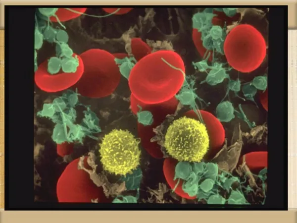
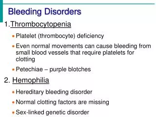
Sites of bleeding in thrombocytopenia • Skin and mucous membranes • Petechiae • Ecchymosis • Hemorrhagic vesicles • Gingival bleeding and epistaxis • Menorrhagia • Gastrointestinal bleeding • Intracranial bleeding
Petechiae Do not blanch with pressure (cf. angiomas)Not palpable (cf. vasculitis)
Quantitative disorders Abnormal distribution Dilution effect Decreased production Increased destruction Qualitative disorders Inherited disorders (rare) Acquired disorders Medications Chronic renal failure Cardiopulmonary bypass Classification of platelet disorders
Associated with bleeding Immune-mediated thrombocytopenia (ITP) Most drug-induced thrombocytopenias Most others Associated with thrombosis Thrombotic thrombocytopenic purpura DIC Trousseau’s syndrome Heparin-associated thrombocytopenia Acquired thrombocytopenia with shortened platelet survival
Approach to the thrombocytopenic patient • History • Is the patient bleeding? • Are there symptoms of a secondary illness? (neoplasm, infection, autoimmune disease) • Is there a history of medications, alcohol use, or recent transfusion? • Are there risk factors for HIV infection? • Is there a family history of thrombocytopenia? • Do the sites of bleeding suggest a platelet defect? • Assess the number and function of platelets • CBC with peripheral smear • Platelet function study
Platelet function screen • Replaces the bleeding time as a test of platelet function • PFA-100; ordered as “platelet function screen” • Blue top tube • Measures the time it takes for blood to block membrane coated with either collagen/epinephrine or collagen/ADP
Platelet function screenResults Epi ADP Interpretation Normal Normal Normal platelet function Abnormal Normal “Aspirin effect” Abnormal Abnormal Abnormal platelet function Valvular heart disease Renal failure Von Willebrand disease
Platelet transfusions • Source • Platelet concentrate (Random donor) Each donor unit should increase platelet count ~10,000 /µl • Pheresis platelets (Single donor) • Storage • Up to 5 days at room temperature • “Platelet trigger” • Bone marrow suppressed patient (>10-20,000/µl) • Bleeding/surgical patient (>50,000/µl)
Platelet transfusions - complications • Transfusion reactions • Higher incidence than in RBC transfusions • Related to length of storage/leukocytes/RBC mismatch • Bacterial contamination • Platelet transfusion refractoriness • Alloimmune destruction of platelets (HLA antigens) • Non-immune refractoriness • Microangiopathic hemolytic anemia • Coagulopathy • Splenic sequestration • Fever and infection • Medications (Amphotericin, vancomycin, ATG, Interferons)
Laboratory Evaluation of BleedingOverview CBC and smear Platelet count Thrombocytopenia RBC and platelet morphology TTP, DIC, etc. Coagulation Prothrombin time Extrinsic/common pathways Partial thromboplastin time Intrinsic/common pathways Coagulation factor assays Specific factor deficiencies 50:50 mix Inhibitors (e.g., antibodies) Fibrinogen assay Decreased fibrinogen Thrombin time Qualitative/quantitative fibrinogen defects FDPs or D-dimer Fibrinolysis (DIC) Platelet function von Willebrand factor vWD Bleeding time In vivo test (non-specific) Platelet function analyzer (PFA) Qualitative platelet disorders and vWD Platelet function tests Qualitative platelet disorders
Adjunctive drug therapy for bleeding • Fresh frozen plasma • Cryoprecipitate • Epsilon-amino-caproic acid (Amicar) • DDAVP • Recombinant human factor VIIa (Novoseven)
Fresh frozen plasma • Content - plasma (decreased factor V and VIII) • Indications • Multiple coagulation deficiencies (liver disease, trauma) • DIC • Warfarin reversal • Coagulation deficiency (factor XI or VII) • Dose (225 ml/unit) • 10-15 ml/kg • Note • Viral screened product • ABO compatible
Cryoprecipitate • Prepared from FFP • Content • Factor VIII, von Willebrand factor, fibrinogen • Indications • Fibrinogen deficiency • Uremia • von Willebrand disease • Dose (1 unit = 1 bag) • 1-2 units/10 kg body weight
Aminocaproic acid (Amicar) • Mechanism • Prevent activation plaminogen -> plasmin • Dose • 50mg/kg po or IV q 4 hr • Uses • Primary menorrhagia • Oral bleeding • Bleeding in patients with thrombocytopenia • Blood loss during cardiac surgery • Side effects • GI toxicity • Thrombi formation
Desmopressin (DDAVP) • Mechanism • Increased release of VWF from endothelium • Dose • 0.3µg/kg IV q12 hrs • 150mg intranasal q12hrs • Uses • Most patients with von Willebrand disease • Mild hemophilia A • Side effects • Facial flushing and headache • Water retention and hyponatremia
Recombinant human factor VIIa(rhVIIa; Novoseven) • Mechanism • Activates coagulation system through extrinsic pathway • Approved Use • Factor VIII inhibitors in hemophiliacs • Dose: (1.2 mg/vial) • 90 µg/kg q 2 hr • “Adjust as clinically indicated” • Cost (70 kg person) @ $1/µg • ~$5,000/dose or $60,000/day
Recombinant human factor VIIain non-approved settings • Surgery or trauma with profuse bleeding • Consider in patients with excessive bleeding without apparent surgical source and no response to other components • Dose: 50-100ug/kg for 1-2 doses • Risk of thrombotic complications not well defined • Anticoagulation therapy with bleeding • 20ug/kg with FFP if life or limb at risk; repeat if needed for bleeding
Approach to bleeding: Summary • Identify and correct any specific defect of hemostasis • Use non-transfusional drugs whenever possible • RBC transfusion for surgical procedures or large blood loss