Download

1 / 30
320 likes | 975 Vues
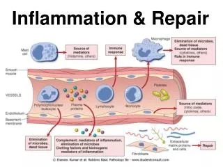
Inflammation and repair. Dr Shaesta Naseem 19-09-13. Inflammation and repair. Inflammatory cells : 1. Macrophages 2. Neutrophils 3. Lymphocytes Etc . . The inflammation. We see. types. Chronic: For long time. Acute: Short time We see neutrophils.

E N D
Inflammation and repair Dr ShaestaNaseem 19-09-13
Inflammation and repair Inflammatory cells : 1. Macrophages 2. Neutrophils 3. Lymphocytes Etc . The inflammation We see types Chronic: For long time Acute: Short time We see neutrophils
CLINICAL FEATURES OF INFLAMMATION : • Tumor (swelling / EDEMA) • Rubor (Redness) • Colar (Warmth / Heat) • Dolor (Pain) • FunctioLasea (loss of function)
1- Fibrinous pericarditis Outer layer of the heart Means/leads to inflammation Fibrinous is found in the blood
The next slide is about this region Organ: Pericardium Dx: Fibrinous Inflammation Acute Means diagnosis
ACUTE FIBRINOUS PERICARDITIS مثل النقاط
ACUTE FIBRINOUS PERICARDITIS غشاء التامور في القلب
FibrinousPericarditis: Section of Heart shows: It becomes thick while the normal is thin • The pericardium is distorted by thick irregular layer of pinkish fibrinous exudate with some red cells and inflammatory cells. • The subpericardiallayer is thickened by edema and shows dilated blood vessels, chronic inflammatory cells and areas of calcification. RBC from blood outside the heart It becomes chronic if it doesn’t treated.
ACUTE APPENDICITIS fat Lumen Here’s the question : What is the white junk coating the surface?
Lumen fills with dead material (inflammatory cells)
Acute appendicitis The same appendicitis but from other side
ACUTE APPENDICITIS Mucosa ulcerated Inflammatory cells
Edema not fat Because it’s not a normal site for fat ACUTE APPENDICITIS neutrophils lymphocytes eosinophil
Acute Suppurative appendicitis:Cross Section of Appendix shows: • On gross, fibrino-purulent exudate is present on the serosal surface. • Accumulation of inflammatory exudate and pus cells in the lumen and the mucosa is ulcerated. • All layers of the appendix wall show edema, dilated and congested blood vessels and infiltration by many neutrophils.
Something related to the hair . collection Has a cavity , opening between two ends . 3-Skin pilonidal sinus Sinus: Track leading Connection between a cavity and epithelial surface Fistula: Abnormal pathway that leads from internal cavity
3- FOREIGN BODY REACTION (PILONIDAL SINUS)
Foreign Body reaction (Pilonidal sinus):Section of Skin Shows: • A sinus tract lined by an inflammatory granulation tissue in the dermis . • The lumen of sinus and wall contain large number of hair shafts with foreign body giant cells, lymphocytes , macrophages & neutrophils .
4-Chronic cholecystitis with stones Means inflammation of gallbladder
Abnormal cavity happens because of the stone CHRONIC CHOLECYSTITIS mucosa submucosa Muscle cells
Chronic cholecystitis:Section of gallbladder wall shows: • Irregular mucosal folds and foci of ulceration in mucosa. • Wall is penetrated by mucosal glands which are present in muscle coat ( Rokitansky- Aschoff sinuses). • All layers show chronic inflammatory cells infiltration and fibrosis. sometimes
5- Brain abscess Liquefactive necrosis
GRANULATION TISSUE young capillaries fibroblast
GRANULATION TISSUE Function : healing
Granulation Tissue:Section of fragments of edematous, loose connective tissue shows: • Many small newly formed capillaries lined by plump endothelial cells. • Proliferation of fibroblasts is seen. • Inflammatory cells including macrophages, lymphocytes, plasma cells and neutrophils in the oedematous stroma. • Pink homogenous collagen fibres may be identified.