CARDIOVASCULAR EXAMINATION
CARDIOVASCULAR EXAMINATION. Steven A. Haist, MD, MS. Division of General Internal Medicine and Geriatrics. Department of Internal Medicine. CARDIOVASCULAR EXAMINATION History Physical Examination Laboratory Tests (CPK, LDH, cholesterol, etc.) Electrocardiography Cardiac imaging—

CARDIOVASCULAR EXAMINATION
E N D
Presentation Transcript
CARDIOVASCULAR EXAMINATION Steven A. Haist, MD, MS Division of General Internal Medicine and Geriatrics Department of Internal Medicine
CARDIOVASCULAR EXAMINATION • History • Physical Examination • Laboratory Tests (CPK, LDH, cholesterol, etc.) • Electrocardiography • Cardiac imaging— • Echocardiography • CT Scan • MRI • Cardiac Catheterization • Nuclear Imaging
CARDIOVASCULAR SYMPTOMS • Chest Pain • Shortness of Breath (dyspnea) • DOE (dyspnea on exertion) • Orthopnea • PND (paroxysmal nocturnal dyspnea) • Trepopnea • Wheezing
CARDIOVASCULAR SYMPTOMS • (continued) • Dizziness / Syncope • Palpitations • Fatigue • Edema • Intermittent claudication • Cyanosis
CHEST PAIN • Angina Pectoris Esophageal Spasm • Myocardial Infarction Cholecystitis • Pericarditis Peptic Ulcer Disease • Pulmonary Embolus Costochondritis • Aortic Dissection Hyperventilation • Esophagitis Mitral Valve Prolapse
HISTORY • Location • Quality • Quantity • Radiation • Timing—Onset, duration, frequency • Setting
HISTORY (continued) • Aggravating Factors • Alleviating Factors • Associated Factors • Pertinent Negatives • Pertinent Past History • Previous Laboratory Tests (prior to this visit) • Risk Factors
HISTORY MYOCARDIAL INFARCTION • Anterior mid-chest (substernal) • Heavy, crushing, pressure-like pain • 9/10 with 10 being the worst pain of their life • Radiates into L arm or neck • > 30 minutes, < 12-24 hours • Awoke this morning with the pain
HISTORY - MI (continued) • Any activity • None • Associated diaphoresis, dyspnea, and nausea • Denies history of MI, murmur, palpitations, orthopnea, DOE,PND • Similar pain not as severe in past lasting 5-10 minutes,relieved with rest, brought on by walking • ECG in ER 1 yr. ago reportedly normal • Smokes 1 PPD, hypertension for 10 years • Father MI age 45, chol 300, no hx DM
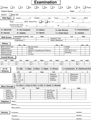
CARDIOVASCULAR PHYSICAL EXAMINATION • General Appearance • Vital Signs • Jugular Veins • Heart • Peripheral Pulses
PHYSICAL EXAMINATION • Is the patient in acute distress? • Always use a hospital gown. Never palpate or auscultate through clothing. • Is the patient comfortable? • Be concerned with the patient's privacy. • Bed at 30° • Must have quiet room ! • Examine from the right side.
Vital Signs • BPboth arms • hypertension • hypotension • orthostatic hypotension • HRtachycardia • bradycardia • Rhythmregular • regularly irregular • irregularly irregular • Respirations tachypnea • Temperature fever
INSPECTION • Jugular veins / jugular venous pressure • Right side, head tilted to L • Adjust angle of bed to see pulsation at mid-neck. • Record distance from R atrium to top of pulsation (sternal angle is 5 cm above RA)
INSPECTION (continued) • Lips, nail beds • Heart: apical impulse • point of maximal impulse • Extremities: (edema, venous or arterial insufficiency)
CARDIAC EXAMINATION • Inspection • Palpation • Percussion • Auscultation
PALPATION • Impulses - finger pads • Thrills (vibrations palpated secondary to a murmur—turbulent blood flow through a heart valve) - Bony part of hand, ball of hand
PALPATION (continued) • Apical impulse (normally 5th ICS and medial to mid-clavicular line) • Point of maximal impulse (PMI) • Left lateral decubitus position (heart closer to chest well) apical impulse more easily palpable
AUSCULTATION • Diaphragm – medium and high frequency sounds • Bell – low frequency sounds • Normally hear closure of valve • Sounds from left side of heart louder than equivalent sounds from right side of heart
AUSCULTATION • S1 – closure of mitral and tricuspid valves • S2 – closure of aortic and pulmonic valves • Low pitched sounds S3, S4, mitral stenosis, and Korotkoff sounds • S1 systole S2 diastole S1 • Simultaneous palpation of carotid pulse can help in differentiating S1 and S2
FIRST AND SECOND HEART SOUNDS • Aortic component (A2) normally louder than pulmonic component (P2) • Mitral component (M1) normally louder than tricuspid component (T1)
FIRST AND SECOND HEART SOUNDS (continued) • T1 and P2 normally heard only over their respective area (LLSB and L2ICS) • Normally left-sided sounds occur first M1T1 (S1) and A2P2 (S2) • S2 changes with respiration, S1 does not Inspiration S1 systole A2 P2 • Expiration S1 systole A2 P2
DIAPHRAGM • Right 2nd intercostal space Aortic Area • Left 2nd intercostal space Pulmonic Area • Third intercostal space Erb’s point • Left lower sternal border Tricuspid area • Apex – over apical impulse Mitral area
BELL • Left lower sternal border • Apex • Apex with patient in left lateral decubitus position • Light pressure only!
POSITIONS • Lying at 30°, standard position • Apex with the patient in the left lateral decubitus position, with bell (mitral stenosis) • At LLSB with patient sitting, leaning forward, fully exhaled with diaphragm(aortic regurgitation)
Observe, record, tabulate, communicate. Use your five senses. The art of the practice of medicine is to be learned only by experience ; 'tis not an inheritance ; it cannot be revealed. Learn to see, learn to hear, learn to feel, learn to smell, and know that by practice alone can you become expert. Medicine is learned by the bedside and not in the classroom. Let not your conceptions of the manifestations of disease come from words heard in the lecture room or read from the book. See, and then reason and compare and control. But see first. No two eyes see the same thing. No two mirrors give forth the same reflection. Let the word be your slave and not your master. Live in the ward. Do not waste the hours of daylight in listening to that which you may read by night. But when you have seen, read. And when you can, read the original descriptions of the masters who, with crude methods of study, saw so clearly. Record that which you have seen ; make a note at the time ; do not wait. * The flighty purpose never is o'ertook, unless the deed go with it.' . . ,1
TERMINOLOGY • Stenosis - forward obstruction • Regurgitation (insufficiency) - backward flow • Aortic Stenosis - during systole forward flow through • obstructed aortic valve from left ventricle • Mitral Stenosis - during diastole forward flow through • obstructed mitral valve from left atrium • Aortic regurgitation - during diastole backward • flow through aortic valve from aorta