
Ca 2+
E N D
Presentation Transcript
C4a C1r C1s C1q b Ca2+ activation stage C4
C2b C1r C1s b C1q Ca2+ C2a Generation of C3-convertase C2 C4a C4b a Mg2+
C1r C5a C3a C1s C1q Ca2+ C2 b a b Generation of C5-convertase C2b C4a C4b2a is C5 convertase Mg2+ C5 C3 C4b
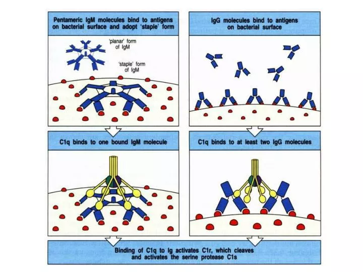
(2)Activation step generation of C3 convertase ----Activated C1s enzymatically cleaves C4 into C4a and C4b. ----Activated C1s enzymatically cleaves C2 into C2a and C2b. ----C4b2b complex binding to the membrane, is known asC3 convertasegeneration of C5 convertase ----C3 convertase cleaves C3 into C3a and C3b. -----C3b binds to the membrane to form C4b2b3b complex. -----C4b2b3b complex functions as C5 convertase
3)Effect step C5 convertase cleaves C5 into C5a and C5b. C5b binds the membrane. C5b binds C6 and C7 to yield a hydrophobic C5b67 complex which attaches quickly to the cell membrane.C8 binds to this complex and causes the insertion of several C9 molecules.C5b6789 complex is known as MAC which leads to formation of a hole in the membrane resulting in cell lysis.
C4 and C2 activation (generation of C3 convertase) Activated C1s enzymatically cleaves C4 into C4a and C4b. C4b binds to the Ag-bearing particle orcell membranewhile C4a remains a biologically active peptide at the reaction site. C4b binds C2 which becomes susceptible to C1s and is cleaved into C2a and C2b. C2b remains complexed with C4b whereas C2a is released in the micro environment. C4b2b complex, is known as C3 convertase in which C2b is the enzymatic moiety.
C3 activation (generation of C5 convertase) C3 convertase, in the presence of Mg,++cleaves C3 into C3a and C3b. C3b binds to the membrane to form C4b2b3b complex whereas C3a remains in the micro environment. C4b2b3b complex functions as C5 convertase which cleaves C5 into C5a and C5b. Generation of C5 convertase marks the end of the classical pathway.
LECTIN PATHWAY C4 activation can be achieved without antibody and C1 participation by the lectin pathway . This pathway is initiated by three proteins: Amannan-bindinglectin(MBL), also known as mannan-binding protein (MBP) which interacts with two mannan-binding lectin-associated serine proteases (MASP and MADSP2), analogous to C1r and C1s. This interaction generates a complex analogous to C1qrs and leads to antibody -independent activation of the classical pathway. C1q can also bind to a number of agents including some retroviruses, mycoplasma, poly-inosinicacid and aggregated IgG, and initiate the classical pathway.
Inosinic acid Inosinic acidorinosine monophosphate (IMP) is anucleosidemonophosphate. Inosinic acid is important inmetabolism. It is theribonucleotideofhypoxanthineand the first nucleotide formed during the synthesis ofpurine. It is formed by thedeaminationofadenosine monophosphate, and ishydrolysedto forminosine. Important derivatives of inosinic acid include purine nucleotides found innucleic acidsandadenosine triphosphate, which is used to storechemical energyinmuscleand other tissues. In the food industry, inosinic acid and itssaltssuch asdisodium inosinateare used asflavour enhancers
مسیر لکتین فعال شدن کمپلمان در غیاب آنتی بادی و بوسیلة اتصال پلی ساکاریدهای میکروبی به لکتین های در گردش مثل MBL است به نظر می رسد فعال شدن کمپلمان از راه لکتینی شبیه راه کلاسیک است MBLبه کربوهیدرات های سطح باکتری ها ی فعال کننده یا سایر مواد متصل شده و متحمل تغییر شکل فضایی مشابه حوادثی که در C1q اتفاق می افتد می گردد
لکتین های دیگری مانند لکتین شناسایی کنندة N استیل گلوکز آمین که بنام فیکولین هستند و از نظر ساختاری مشابه c1q می باشند از یک طرف به پلی ساکاریدهای میکروبی متصل شده و از طرف دیگر به سرین پروتئازهای همراه MBL یا (MASPs) ها از نوع 1و 2و3 نیز متصل می شوند الیگو مرهای بلندتر MBL با MASP-2 و MASP-3 همراه می شوند این دو پروتئاز یک کمپلکس مشابه با کمپلکس تشکیل شده بوسیله c1r و C1s را تشکیل می دهند و MASP-1مولکول های C4 و C2 را می شکند
Ficolin A binding protein of 40 kDa forTGF-beta-1isolated originally fromporcineuterus membranes Two closely relatedcDNAclones encode proteins composed mainly of fibrinogen-like and collagen-likedomains The corresponding proteins are calledFicolin-alphaandFicolin-beta Ficolins have a similar overall structure to complement component C1q and the collectins
lectin pathway
پروپردين پروتئين هايي در سرم بنام پروپردين وجود دارد که در مجاورت مادة زيموزان که پلي ساکاريدي در ديوارة سلولي مخمر است مي توانند سيستم کمپلمان را فعال نمايند
alternative pathway activator LPS bacteria zymosan dextran IgA IgG4 IgE
ALTERNATIVE PATHWAY Alternative pathway begins with the activation of C3 and requires Factors B and D and Mg++ cation, all present in normal serum. Spontaneous activation of C3A metastable C3b-like molecule (C3i) is generated by slow hydrolysis of the native C3. C3i binds factor B which is cleaved by Factor D to produce C3iBb. C3iBb complex cleaves native C3 into C3a and C3b. C3b binds factor B, which is again cleaved by Factor D to produce C3bBb(C3 convertase) This C3 convertase (or the one generated by classical pathway: (C4b2a), if not inactivated, will continue to act on C3 and cause its total depletion.
مولکول C3i اولين جزء لازم براي فعال شدن راه آلترناتيو قطعة C3b است که به مقدار جزئي و بطور مداوم در جريان خون بوجود مي آيد مولکول C3 در جريان خون به دو صورت ديده مي شود ، يکي شکل طبيعي و دست نخورده که حاوي پيوندهاي تيواستر است و ديگري شکل تغيير يافته که به صورت C3(H2O) يا C3i نشان داده مي شود که در آن پيوندهاي تيواستر توسط آب هيدروليز شده است C3 طبيعي در پلاسما و بطور مداوم و به مقدار کم هيدروليز شده و پيوندهاي تيواستر داخلي آن تغيير يافته و مولکول C3i توليد مي شود
C3bBb کونورتاز مولکول C3i در مجاورت يون منيزيوم با پروتئيني بنام فاکتور B تشكيل كمپلکس (C3iBF) را مي دهد پروتئين ديگري بنام فاکتور D که خاصيت آنزيمي دارد فاکتور B را در اين مجموعه شکسته و به دو جزء Ba و Bb تبديل مي کند قطعة بزرگتر (Bb) با C3i تشــــکيل کمپلکس آنزيمي را مي دهد که خود يک آنزيم تبديل کننده بوده و مي تواند C3 را به اجزاء C3a و C3b تبديل کند برخي از مولکول هاي C3b که به اين ترتيب بوجود مي آيند در مجاورت کمپلکس C3iBb بر سطح آنتی ژن فرود آمده و سپس فاکتور B بر اين مولکول هاي C3b تأثير کرده و به علت پايداري بيشتر به سمت تشکيل کمپلکس پيش مي رود اين کمپلکس قادر است تعداد بيشتري مولکول C3 را به C3b و C3a تبديل کند بنابراین کمپلکس C3bBb کونورتاز مسیر آلترنایو است و عملش شکستن مولکول های C3 بیشتر است
C3-Convertase حتی اگر C3b بوسیلة مسیر کلاسیک تولید شود می تواند با Bb کمپلکس تشکیل داده و این کمپلکس قادر به شکستن مولکول های C3 بیشتری است بنابراین C3 کونورتاز مسیر آلترناتیو وقتی که بوسیلة مسیر آلترناتیو یا کلاسیک یا لکتین فعال شده باشد ، فعالیت کمپلمان را تقویت می کند اما يک مجتمع ناپايدار بوده و به مرور تجزيه مي شود مگر اينکه پروتئيني بنام فاکتور پروپردين (P) به اين مجموعه اضافه شود فاکتور P پلاسما به مجموعه قبلي اضافه شده و تشکيل کمپلکس پايدار را مي دهد که مي تواند بخوبي سبب تجزيه مولکول هاي C3 گردد به همين علت اين مولکول را تبديل کنندة C3 يا (C3-Convertase) در مسير آلترناتيو مي ناميم
حال مقادير بيشتري از پروتئين C3 تجزيه شده و به صورت يک حلقة خود تنظيم مثبت مقادير بيشتري از آنزيم C3 کانورتاز را تشکيل ميدهد بنابراين با بوجود آمدن مولکول هاي بيشتر C3b و اتصال به C3 کانورتاز ، کمپلکس هائي از تشکيل مي گردد که خاصيت تبديل کنندگي مولکول C5 را داشته و بنام C5-Convertase ناميده مي شود C5-Convertase پروتئين C5 را به دو جزء C5a و C5b تبديل مي کند ، با بوجود آمدن جزء C5b اولين جزء در تشکيل کمپلکس حمله غشائي ايجاد شده و هر دو مسير کلاسيک و آلترناتيو به يکديگر مي پيوندد
شباهت هائ دو مسیر پروتئین ها از نظر خواص فيزيکي و شيميايي و مکانيزم فعال شدن مشابهت دارند آنزیم هاي C1s و فاکتور D شبيه به يکديگرند زيرا هر دو پروتئين از آنزیم هاي سرين استرازي مي باشند آنزيم C1sپروتئین ها ي C4 و C2 را مي شکند که سرانجام آنزيم جديد C4b2b يا C3 کانورتاز درست مي شود فاکتورD هم در مجاورت قطعة C3b فاکتور B را که از نظر فيزيکي و شيميايي شباهت بسياري با پروتئين C2 دارد شکسته و در مجاورت يون منيزيوم مجموعاً تشکيل آنزيم C3bBbP ياC3 کانورتاز را مي دهد هر دو آنزيم C3 کانورتاز يک پيوند پپتيدي را در مولکول C3 شکسته و با قطعة C3b مجموعاً آنزيم جديدي را بوجود مي آورند آنزیم هایC4b2b3b در مسير کلاسيک و (C3b)nBbP در مسير آلترناتيو پروتئين C5 را مي شکنند و بنابراين C5 کانورتاز ناميده مي شوند
Normal regulation of C3 convertase C3b, in fluid phase, is very short lived unless it finds a suitable stabilizing membrane or molecule (C3 activator). In the absence of exogenous pathogen, it binds quickly to autologous red cells via the C3b receptor, CR1 at a site close to decay accelerating factor (DAF) which prevents the binding of Factor B. Binding to CR1 also makes C3b susceptible to Factor I which cleaves it into many fragments (iC3b, C3d, C3e, etc) .
C4b, generated in the classical pathway, is also regulated by DAF, CR1 and Factor I. A defect in or deficiency of DAF can lead to cell lysis and anemia, as in its absence further activation of C will proceed and lead to the membrane attack pathway and cell lysis.
Another serum protein, factor H, can displace factor B and bind to C3b. Binding of factor H makes C3b more susceptible to factor I. C3 convertase generated by the classical pathway is regulated also in a similar manner by DAF, Cr1 and Factor I. The only difference is that C4b-binding protein (C4b-BP, not factor H) makes it susceptible to Factor I. A genetic deficiency of factor I (or factor H) leads to uncontrolled C3 activation and is a major cause of inherited C3 deficiency
Stabilization of C3 convertase Certain bacteria or their products (peptidoglycan, polysaccharides, etc.), provide a protected (activator) surface for C3b. Thus, C3b bound to such a surface is relatively resistant to the action of factor I. Even membrane bound C3bBb dissociates fairly rapidly. However, binding of another protein, properdin, further stabilizes this complex. It is for this reason, the alternative pathway is also called the properdin pathway.
Generation of C5 convertase Stabilized C3 convertase cleaves more C3 and produces C3bBbC3b complex (analogous to C4b2a3b of the classical pathway), the C5 convertase which cleaves C5 into C5a and C5b. C5b initiates the membrane attack pathway which leads to cell lysis. While these pathways of C3 activation are initiated by different mechanisms, they are analogous to each other and both can lead to membrane lysis. The alternative pathway provides a means of non-specific resistance against infection without the participation of antibodies and hence provides a first line of defense against a number of infectious agents.
Manygram negativeand somegram positivebacteria, Certain viruses, Parasites, Heterologous red cells, Aggregated immunoglobulins (particularly, IgA), Some other proteins (e.g. proteases, clotting pathway products) can activate the alternative pathway. One protein, cobra venom factor (CVF), has been extensively studied for its ability to activate this pathway
LYTIC PATHWAY The lytic (membrane attack) pathway involves the C5-9 components C5 convertase generated by the classical or alternative pathway cleaves C5 into C5a and C5b. C5b binds C6 and subsequently C7 to yield a hydrophobic C5b67 complex which attaches quickly to the plasma membrane. Subsequently, C8 binds to this complex and causes the insertion of several C9 molecules,bind to this complex and lead to formation of a hole in the membrane resulting in cell lysis. The lysis of target cell by C5b6789 complex is nonenzymatic and is believed to be due to a physical change in the plasma membrane. C5b67 can bind indiscriminately(randomly) to any cell membrane leading to cell lysis. Such an indiscriminate damage to by-standing cells is prevented by protein S (vitronectin) which binds to C5b67 complex and blocks its indiscriminate binding to cells other than the primary target.
BIOLOGICALLY ACTIVE PRODUCTS OF COMPLEMENT ACTIVATION Activation of complement results in the production of several biologically active molecules which contribute to resistance, anaphylaxis inflammation. Kininproduction C2b generated during the classical pathway of C activation is a prokinin which becomes biologically active following enzymatic alteration by plasmin.
Excess C2b production is prevented by limiting C2 activation by C1 inhibitor (C1-INH) also known as serpin which displaces C1rs from the C1qrs complex. A genetic deficiency of C1-INH results in an overproduction of C2b and is the cause of hereditary angioneurotic edema. This condition can be treated withDanazolwhich promotes C1-INH production or with ε-amino caproic acid which decreases plasmin activity.
Anaphylotoxins C4a, C3a and C5a (in increasing order of activity) are all Anaphylotoxins which cause basophil/mast cell degranulation and smooth muscle contraction. Undesirable effects of these peptides are controlled by carboxypeptidase B (C3a-INA). Chemotactic Factors C5a and MAC (C5b67) are both chemotactic. C5a is also a potent activator of neutrophils, basophils and macrophages and causes induction of adhesion molecules on vascular endothelial cells. Opsonins C3b and C4b in the surface of microorganisms attach to C-receptor (CR1) on phagocytic cells and promote phagocytosis
Other Biologically active products of C activation Degradation products of C3 (iC3b, C3d and C3e) also bind to different cells by distinct receptors and modulate their functions In summary, the complement system takes part in both specific and non-specific resistance and generates a number of products of biological and pathophysiological significance (Table 2). There are known genetic deficiencies of most individual C complement components, but C3 deficiency is most serious and fatal. Complement deficiencies also occur in immune complex diseases(e.g.SLE) and acute and chronic bacterial, viral and parasitic infections.