Download
1 / 74
760 likes | 1.06k Vues
Neck Region. PA 481. Anterior Surface. Triangles at Surface. Hyoid. C3 level Suprahyoid muscles Infrahyoid muscles. Vertebral Column. Intervertebral disc. Vertebra Parts. C1 & C2. Cervical Vertebra. Typical Cervical Vertebra. Spinal Cord.

E N D
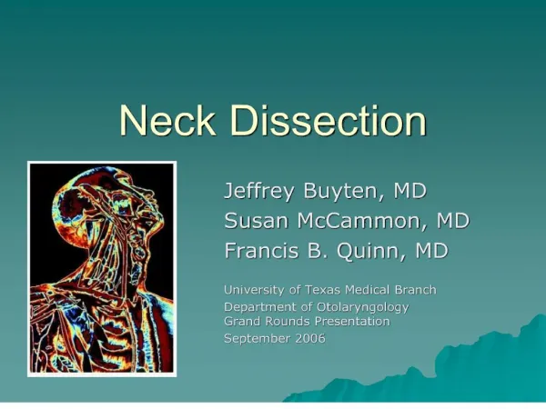
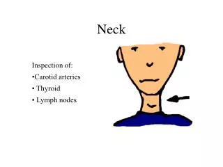
Neck Region PA 481
Hyoid • C3 level • Suprahyoid muscles • Infrahyoid muscles
Spinal Cord • Receives and generates signals to body through the spinal nerves
Cord in Spinal Canal Posterior Median Sulcus Posterior Root Denticulate Ligament Dorsal Root Ganglion Anterior Root Spinal Nerve
Functional Arrangement of SC Ascending and descending pathways
Cervical Plexus Serves neck and diaphragm
Brachial Plexus Innervates most of the arm and someof the body wall.
Spinal Cord Segments • 4 segments: Cervical, Thoracic, Lumbar, and Sacral (only 1 coccygeal nerve) • 31 pairs of spinal nerves
Neck Fascia Superficial fascia Prevertebral fascia Alar fascia Investing fascia Pretracheal fascia
SCM and Trapezius SCM Trapezius
Cervical Triangles Anterior Triangle Posterior Triangle
Posterior Triangle Occipital triangle Supraclavicular triangle
Superficial and Deep Posterior Triangle Ext. jugular v. SCM Splenius Levator scapulae Nerve Point of Neck Accessory n. Phrenic n. Prevertebral fascia Platysma Scalenes Brachial plexus
Supraclavicular Triangle Brachial Plexus SCM Subclavian vessels
Anterior Triangles Submandibular Submental Carotid Muscular
Muscles of Triangle Thyrohyoid Sternothyroid Thyroid Sternohyoid
Accessory Thyroid gland Accessory thyroid along thyroglossal duct
Pyramidal lobe (50% of people have this lobe structure)
Thyroid Follicle (follicular cells thyroxine) Parafollicular cells calcitonin
Control of Thyroxine Secretion Short loop Long loop
Thyroid Malfunction • Hypothyroidism • Endemic goiters –due to iodine deffeicency • Cretinism –i thyroxine in child results in igrowth (dwarf) and severe mental retardation • Myxedema –i thyroxine in adult, leads to swelling of tissues plus other symptoms
Thyroid Malfunction • Hyperthyroidism • Toxic goiters (Graves disease) –Ab may stimulate thyroid without negative feedback control • Exophthalmos –symptom present in many hyperthyroid patients
PTH Actions • Stimulates resorption of bone hCa+ and PO4- in blood • Stimulates Ca+ absorption in intestine (active Vit. D3 necessary for Ca+ absorption) • Stimulates Ca+ reabsorption and PO4- excretion in kidney • Stimulates Vit. D3 formation (skin) and activation (kidney) • Vital for life
Laryngeal Cartilages Hyoid Epiglottis (anterior) Arytenoid cartilage Thyroid cartilage Cricoid cartilage (posterior)
Voice Box Vestibular fold Ventricle Vocal fold
CN X: supply to laryngeal muscles Inf. Vagal ganglion Internal Laryngeal n.(sensory & autonomic) External Laryngeal n.(motor to inf. Pharnygeal constrictor and the cricohyoid) Recurrent Laryngeal n.(motor to all other laryngeal muscles)