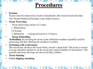
Procedures
Procedures. Basic Format ORIF Femur (Femoral Shaft) Level II. Objectives. Assess the anatomy, physiology, and pathophysiology of the ORIF Femur. Analyze the diagnostic and surgical interventions for a patient undergoing an ORIF Femur.

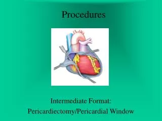
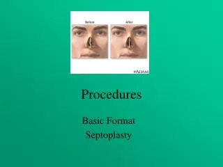
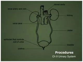
Procedures
E N D
Presentation Transcript
Procedures Basic Format ORIF Femur (Femoral Shaft) Level II
Objectives • Assess the anatomy, physiology, and pathophysiology of the ORIF Femur. • Analyze the diagnostic and surgical interventions for a patient undergoing an ORIF Femur. • Plan the intraoperative course for a patient undergoing ORIF Femur. • Assemble supplies, equipment, and instrumentation needed for the procedure.
Objectives • Choose the appropriate patient position • Identify the incision used for the procedure • Analyze the procedural steps for ORIF Femur. • Describe the care of the specimen • Discuss the postoperative considerations for a patient undergoing ORIF Femur .
Terms and Definitions • Open Reduction Internal Fixation • Fixation Devices • Intermedullary Rodding • DHS System (Dynamic Hip Screw System)
Definition/Purpose of Procedure • Realignment and fixation of a fracture of the femur through an operative incision • Goal: solid union of bone in perfect alignment, to return joints and muscles to normal position, to prevent or repair vascular trauma, and to rehabilitate the pt as early as possible
Pathophysiology • Classifications of fractures • Traumatic • Closed (Simple) • Open (Compound) • Pathologic
Pathophysiology: Type of Fractures • Oblique (45 degree angle to bone) • Spiral (curves around bone) • Avulsed (fracture pulls bone & other tissues away from the point of attachment. • Comminuted (bone breaks in many pieces) • Compressed (bone is crushed) • Impacted (broken bone ends are forced into each other) • Depressed (broken bone is forced inward) • Complete vs Incomplete • Stable vs Unstable
Fractures Overview Methods of Treatment: Closed reduction w/immobilization Skeletal traction External fixation Internal fixation Electrostimulation
Internal Fixation • Overview • Screws, Plates, Nails • Intermedullary nailing • Advantages: increase and evenly spread load sharing of the bone, reduced scarring, minimal blood loss, low infection rate, fracture hematoma preserved at fracture site. • Type of nail
Fixation of a short oblique fracture using a plate and screws above and below the fracture
Fixation of a long oblique fracture using screws through the fracture site
Diagnostics • Exams • X-ray • Preoperative Testing
Surgical Intervention:Special Considerations • Patient Factors • Room Set-up • Position sterile tables on the affected side • Etc • Have x-rays in room • Some surgeons prefer to double-glove and remove the first pair following completion of draping • Observe x-ray precautions; notify radiology dept when pt is being positioned for preliminary films; intraop x-ray will be used (either via portable machine or C-Arm flouroscopic image intensification) • Wear protective goggles
Surgical Intervention: Anesthesia • Method: General or epidural • Equipment • Upper body Bair hugger available
Surgical Intervention: Positioning • Position during procedure • Lateral position on Fracture Table or without; might be supine • Supplies and equipment • If no FX Table, may use a leg holder and pillow placed between legs and clear adhesive drape used to drape perineum out of field before skin prep • Wide adhesive to stabilize patient at shoulders in addition to safety strap over the unaffected thigh • Special considerations: high risk areas • Arm on unaffected side on armboard, other arm either flexed across chest or postioned on pillow on Mayo stand • Padding around feet, ankles, bony prominences
Surgical Intervention: Skin Prep • Method of hair removal: wet prep • Anatomic perimeters • Begin over fracture site extending from umbilicus to below the knee, well beyond midline anteriorly, and down table posteriorly. If no FX Table, leg and foot are completely prepared. • Solution options: Betadine, Duraprep, Hibiclens
Surgical Intervention: Draping/Incision • Types of drapes • Folded towels and sterile, plastic adhesive drape. fenestrated sheet covers the field • Order of draping • Four sheets may be draped around perimeter of site; • Special considerations • State/Describe incision: at fracture site
Surgical Intervention: Supplies • General • Basin set, Suction tubing, ESU pencil/holder, needle magnet or counter, asepto or bulb syringe, graduated cylinder, 6 “ tube or impervious stockinette (if FX Table is not used) • Specific • Sterile plastic adhesive U-drapes (2) (perineum and operative site) • Suture & Blades: Blades (2) # 10 • Medications on field (name & purpose) • Catheters & Drains
Surgical Intervention: Instruments • General • Basic orthopedic procedures tray • Specific • AO femoral nail instrumentation set • Hip retractor tray • Bone holding instrumentstray • Drill bits, including ¼ “ gauge • Power reamer and drill and cords, long guide wires, Extra guide pins, screw set • Fixation device: Rods or nails (eg. Hanson-Street, Kuntscher, Rush) or compression set and instrumentation particular to that device
Surgical Intervention: Instruments • Specific • 13-mm calibrated drill bit, Large reverse awl, tissue protector, radiographic ruler, a guide rod for cannulated nails, calibrated wire guide
Kuntschner Nail Examples of Intramedullary nail and rod devices.
Surgical Intervention: Equipment • General • Suction, ESU, Power Source for drill • Specific • Fracture Table or leg holder (optional)
Surgical Intervention: Procedure Steps • Phases of fracture treatment • Reduction • Immobilization • Rehabilitation
Surgical Intervention: Procedure Steps • Surgeon confirms nail length with flouroscopy • Obtains AP view of proximal femur • Ruler is held along the lateral side of the thigh and placed until the top is level with tip of greater trochanter, skin marked at this level • AP view of distal femur is taken • Proximal end of the ruler is placed at the skin mark and nail length is read from the ruler
Surgical Intervention: Procedure Steps • Surgeon makes a longitudinal incision proximal to the greater trochanter thru the gluteus medius and maximus • The 3.2 calibrated guidewire is placed using the power drill. It is placed in the medullary canal to a depth of 100 mm at the entry point where the nail will be placed. • The 13-mm cannulated drill bit with tissue protector is placed over the guidewire and drilled to a depth of 100 mm. This opening will allow for insertion of 9 to 12 mm nails. • For 13- to 15- mm nails, the broach is used instead to enlarge the opening. The drill bit is removed after the opening has been created. • STSR assembles insertion instruments
Surgical Intervention: Procedure Steps • Use of cannulated screws: • The correct connecting screw is placed into the insertion handle and secured to the nail with the ball hexagonal screwdriver. • The driving cap is screwed onto the insertion handle. This serves as the striking point when the hammer is used. • STSR hands the assembly to the surgeon, who manually inserts the nail into the femoral opening as far as possible. • For cannulated nails, the nail is inserted over the guidewire, which passes thru an opening in the side of the insertion handle
Surgical Intervention: Procedure • Next: the surgeon is given the hammer to drive the nail into the distal metaphysis. The guidewire is removed. • Next: the proximal locking bolts are placed: • The standard aiming arm is attached to the insertion handle. The STSR assembles the triple trocar assembly, 11.0/8.0 mm protection sleeve, 8.0/4.0 mm drill sleeve, and 4.00 mm trocar. • The STSR loads the 4.0-mm calibrated drill bit onto the power drill and hands it to the surgeon, who drills thru both cortices until the drill sleeve presses against the cortex. • The locking bolt length is read from the calibrated drill and communicated by the surgeon to the STSR.
Surgical Intervention: Procedure • The STSR hands the surgeon the locking bolt, protection sleeve, and screwdriver to insert the locking bolt. This step is repeated for the second proximal locking bolt. • Next: distal locking bolt is placed with the use of flouroscopy: C-Arm is placed to show the most distal hole in the femoral nail • A stab incision is made over the site of the distal hole with a # 10 knife blade • STSR hands surgeon the power drill w/4.0 mm drill bit, which is placed in the distal hole and drilled thru both cortices. • Using a depth gauge, the surgeon measures the length of the locking bolt. • STSR should remind surgeon to add 2-4 mm to the reading to make sure the thread of the bolt engages far into the cortex. • STSR hands the surgeon the correct size bolt, holding sleeve, and screwdriver for insertion of the bolt.
Surgical Intervention: Procedure • Next: insertion instruments are removed. • STSR hands the surgeon the hexagonal screwdriver and end cap to be threaded onto the proximal end of the nail. • STSR should be prepared to hand the surgeon the ratchet wrench to tighten the end cap. • Surgeon closes the stab incisions, places dressings, and pt taken to PACU.
Counts • Initial: Sponges and sharps • First closing • Final closing • Sponges • Sharps
Dressing, Casting, Immobilizers, Etc. • Types & sizes: N/A • Type of tape or method of securing: heavy gauze pad and tape of choice—foam or silk tape
Specimen & Care • Identified as (N/A) • Handled: routine, etc.
Postoperative Care • Destination • PACU • Expected prognosis (Good, Depends on Dx)
Postoperative Care • Potential complications • Hemorrhage • Infection • Other: Damage to…. • Surgical wound classification: Class I, unless traumatic injury with visible debris--
Resources • Alexander’s pp. 880-882 • Berry & Kohn p. 751- 755 • Goldman pp. 330-332 • Lemone & Burke pp. 1193-1203 • MAVCC Info Sheets Unit 10 • STST pp. 851-853.
Cast Application Supplies and equipment Types Technique
Casting Supplies • Plaster of Paris or fiberglass cast material • Webril and soft roll as needed • Bucket and water to requested temperature • Clean gloves • Cast cutter, spreaders, heavy bandage scissors on standby
Types of Casts: See STST Table 21-5 • Short arm • Long arm • Shoulder spica • Short leg • Long leg • Cylinder • One-and-one half hip spica cast • Body jacket