Gel
&. Gel. Plasmid. Electrophoresis. Mapping. Plasmid Mapping. Purpose: identifying the position of “restriction sites” on a fragment of DNA Can help identify the position of a specific gene within DNA Restriction enzymes (endonuclease) cut a plasmid into smaller fragments of DNA.

Gel
E N D
Presentation Transcript
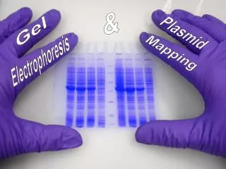
& Gel Plasmid Electrophoresis Mapping
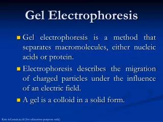
Plasmid Mapping • Purpose: identifying the position of “restriction sites” on a fragment of DNA • Can help identify the position of a specific gene within DNA • Restriction enzymes (endonuclease) cut a plasmid into smaller fragments of DNA
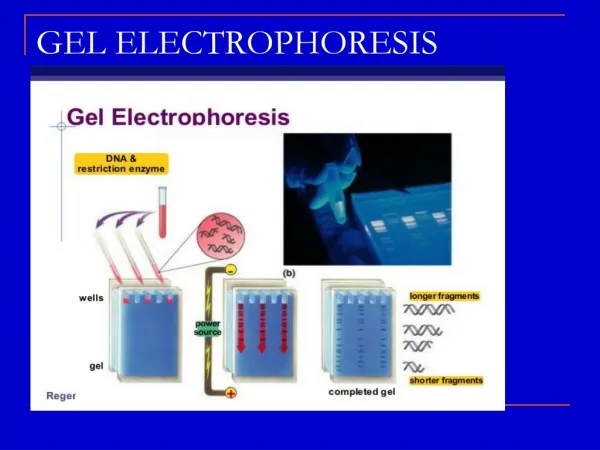
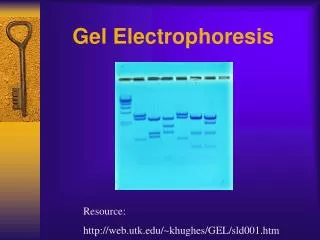
Gel Electrophoresis • Procedure used to separate the DNA fragments by their size
Gel Electrophoresis • Restriction enzymes “cut up” the DNA samples into fragments • DNA samples placed into wells at one end of the chamber • An electric current is applied to the gel – smaller fragments move towards the positive end faster than larger fragments
Example • This fragment of DNA is 7.0 kb (kilobases) in length • When digested with Hind III enzyme, two fragments result (a 6.2 kb fragment and a 0.8 kb fragment)
Example • Thus, we know there is a Hind III restriction site 0.8 kb from one end of the fragment
Example • If we digest the fragment with another enzyme, Sal I, two fragments result • Now, they are 5.8 kb and 1.2 kb in length
Example • If the restriction sites for both enzymes are on the same end, we’d expect the 0.8 kb fragment to be within the 1.2 kb fragment
Example • A double-enzyme digestion would give three fragments (0.4, 0.8, and 5.8 kb)
Example • However, if the restriction sites are at opposite ends, we’d expect the 0.8 kb fragment to be within the 5.8 kb fragment
Example • A double-enzyme digestion would give three fragments (0.8, 5.0, and 1.2 kb)
Example • In order to determine which map is correct, we must digest the DNA with both enzymes