SWI-informed Diffusion Tensor Tractography
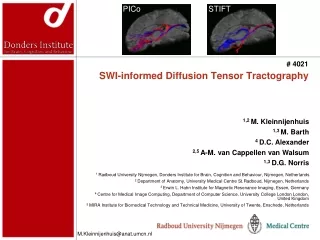
# 4021. PICo. STIFT. SWI-informed Diffusion Tensor Tractography. 1,2 M. Kleinnijenhuis 1,3 M. Barth 4 D.C. Alexander 2,5 A-M. van Cappellen van Walsum 1,3 D.G. Norris. 1 Radboud University Nijmegen, Donders Institute for Brain, Cognition and Behaviour, Nijmegen, Netherlands

SWI-informed Diffusion Tensor Tractography
E N D
Presentation Transcript
# 4021 PICo STIFT SWI-informed Diffusion Tensor Tractography 1,2 M. Kleinnijenhuis 1,3 M. Barth 4 D.C. Alexander 2,5 A-M. van Cappellen van Walsum 1,3 D.G. Norris 1 Radboud University Nijmegen, Donders Institute for Brain, Cognition and Behaviour, Nijmegen, Netherlands 2 Department of Anatomy, University Medical Centre St.Radboud, Nijmegen, Netherlands 3 Erwin L. Hahn Institute for Magnetic Resonance Imaging, Essen, Germany 4 Centre for Medical Image Computing, Department of Computer Science, University College London London, United Kingdom 5 MIRA Institute for Biomedical Technology and Technical Medicine, University of Twente, Enschede, Netherlands M.Kleinnijenhuis@anat.umcn.nl
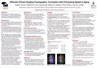
SWI-informed Diffusion Tensor Tractography M.Kleinnijenhuis@anat.umcn.nl # 4021 Problem description Fibre tractography in diffusion weighted images (DWI) suffers from partial volume effects • Typical DWI voxels (8 ml) contain multiple tracts at e.g. tract borders (Fig.1a) • High resolution (0.125 ml) long TE gradient echo images (GRE) also show contrast within the white matter at high field1(Fig.1b) Fig1a. FA Tractography can benefit from combining the diffusion tensor with information from high resolution volumes Fig1b. GRE 1 Li et al., NI 2006
Structure Tensor Informed Fibre Tractography (STIFT) SWI-informed Diffusion Tensor Tractography M.Kleinnijenhuis@anat.umcn.nl # 4021 The structure tensor (ST) is a suitable representation of scalar images to incorporate in tractography algorithms • The ST captures local image features; the principal structure direction (PSD) is given by the first eigenvector of the ST (Fig.2) • In anisotropic diffusion filtering the ST is used to enhance certain features of the image • The ST calculated by edge-enhancing diffusion1 is most useful for enhancing the sheet-like fiber bundles in the GRE image The PSD at tract borders in GRE images is expected to be orthogonal to the principal diffusion direction (PDD) (Fig.2) Fig2. PDD (red) & PSD (green) 1 Weickert, PhD thesis 1996
Fibre tracking with Camino PICo1 and STIFT SWI-informed Diffusion Tensor Tractography M.Kleinnijenhuis@anat.umcn.nl # 4021 • Adapted PICo informed by the structure tensor • Tracking direction (TD) is found by rotating the PDD towards the plane orthogonal to the PSD proportional to its normalized first eigenvalue • The adapted tracking direction is used in white matter only (Eq.1) • Seed point pairs were placed in the GRE image in adjacent voxels within and outside conspicuous fiber bundles Fig3. Tracking Direction 1 Parker et al., JMRI 2003
Optic radiation (OR) Fig4a. PICo NB inferior view Fig4b. STIFT NB inferior view Meyer’s loop V1 extra striate V1 Fig5. Seeds Inside OR Outside OR ∆x = 0.5 mm PICo: • Frontal1 and temporal2 to V1 STIFT: • Meyer’s loop to V13 • Frontal1 and temporal2 to extrastriate areas SWI-informed Diffusion Tensor Tractography M.Kleinnijenhuis@anat.umcn.nl # 4021 1 Inferior occipito-frontal fasciculus (IOFF)) 2 Inferior longitudinal fasciculus (ILF) 3 Optic radiation (OR)
Tapetum (TM) Fig6b. STIFT Fig6a. PICo PICo: • Frontal to V1; mixed for both seeds • Tapetum corpus callosum specific to seed in TM STIFT: • Separation of frontal fibers • Temporal tapetum specific to seed in TM Fig7. Seeds Inside TM Outside TM ∆x = 0.7 mm SWI-informed Diffusion Tensor Tractography M.Kleinnijenhuis@anat.umcn.nl # 4021
Anatomical specificity increased with STIFT SWI-informed Diffusion Tensor Tractography M.Kleinnijenhuis@anat.umcn.nl # 4021 Closely spaced seed points in neighbouring tracts result in well separated tracts using STIFT • From the seedpoint within the OR3, the geniculostriate pathway was tracked, while seeding just outside the OR reconstructed the associative fibers of the IOFF1 and ILF2; PICo tracked the IOFF and ILF to V1 • Different parts of the tapetum were tracked by PICo and STIFT; STIFT showed clearly separate fiber tracts for both seeds Large veins and iron-rich subcortical structures can affect STIFT results negatively At specific locations STIFT can be a valuable tool to increase specificity and accuracy of fiber tracking 1 Inferior occipito-frontal fasciculus (IOFF) 2 Inferior longitudinal fasciculus (ILF) 3 Optic radiation (OR)
Acknowledgements SWI-informed Diffusion Tensor Tractography M.Kleinnijenhuis@anat.umcn.nl # 4021 Thanks to Dirk-Jan Kroon for the implementation of the edge-enhancing diffusion filter
Additional material SWI-informed Diffusion Tensor Tractography M.Kleinnijenhuis@anat.umcn.nl # 4021
Methods: acquisition SWI-informed Diffusion Tensor Tractography M.Kleinnijenhuis@anat.umcn.nl # 4021 DWI, GRE, and T1-weighted images in two healthy volunteers:
Methods: preprocessing (1) SWI-informed Diffusion Tensor Tractography M.Kleinnijenhuis@anat.umcn.nl # 4021 • DWI artefact detection & realignment with PATCH1 • Brain extractionwith FSL • Bias field correction with FSL • Coregistration • GRE T1;with FSL using FAST-based weighting volumes • Mean b=0 DWI T1;with constrained warping2 • GRE image structure tensor with edge-enhancing diffusion3 • Vessel enhancing diffusion with VED4 • WM-GM Segmentation with FSL 1 Zwiers, ISMRM 2009 2 Visser, ISMRM 2010; Poster #3459 3 Kroon & Slump, IEEE-EMBS Benelux 2009 4 Koopmans, MRMP 2008
Methods: preprocessing (2) SWI-informed Diffusion Tensor Tractography M.Kleinnijenhuis@anat.umcn.nl # 4021 Coregistration • GRE T1;with FSL using FAST-based weighting volumes • Mean b=0 DWI T1;with constrained warping in PE direction2 2 Visser, ISMRM 2010; Poster #3459
Methods: preprocessing (3) SWI-informed Diffusion Tensor Tractography M.Kleinnijenhuis@anat.umcn.nl # 4021 GRE image structure tensor with edge-enhancing diffusion3 3 Kroon & Slump, IEEE-EMBS Benelux 2009