STROKE
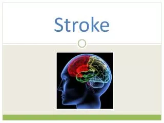
STROKE. Nursing Assessment of the Stroke P atient. STROKE. A stroke , also known as a cerebrovascular accident ( CVA ), is the rapidly developing loss of brain function(s) due to a disturbance in the blood supply to the brain.

STROKE
E N D
Presentation Transcript
STROKE Nursing Assessment of the Stroke Patient.
STROKE A stroke, also known as a cerebrovascular accident (CVA), is the rapidly developing loss of brain function(s) due to a disturbance in the blood supply to the brain. A stroke can be ischemic (lack of blood flow) caused by a blockage (thrombosis, arterial embolism), or haemorrhagic (leakage of blood).As a result, the affected area of the brain is unable to function. Depending which area of the brain is affected, this may result in an inability to move one or more limbs on one side of the body, inability to understand or formulate speech, or an inability to see one side of the visual field. A stroke is a medical emergency and can cause permanent neurological damage, complications, and death.
THE BRAIN • 2% of total body weight • Protected by hard, rigid skull • Three major divisions: - Cerebrum- (2 hemispheres, 4 lobes) • Sensory interpretation, movement control, memory, emotions, personality, reasoning, intelligence etc. - Cerebellum • Coordination of movement and posture, maintains equilibrium with the body - Brain stem- • Connects spinal cord to cerebrum. Vital functions (BP, HR, breathing) and origin of cranial nerves
ICP – Intracranial Pressure • The skull is a rigid compartment which is completely full with essentially non-compressible contents: Brain matter 80%, Blood 10%, CSF 10% • The volume of these contents are in a constant state of equilibrium • ICP is the pressure exerted by the CSF within the intracranial space/skull • Normal ICP= 0-15mmHg • ICP rises during various activities such as coughing, exercise and straining. • Some compensatory mechanisms exist to maintain a constant state of equilibrium within the intracranial space –displacement of CSF, Compression of Dural sinuses and venous system, decrease in the production of CSF and cerebral vasoconstriction. • These compensatory mechanisms are extremely limited. If ICP is elevated for an extended period mortality and morbidity are dramatically increased. • A rise in ICP can be due to a number of illnesses, diseases and accidents: • Head trauma • Cancer or space occupying lesion • Meningitis • SAH • Stroke • Generalized brain swelling (encephalopathy, liver failure) • Hydrocephalus • Hyperthermia • Hypoxia
SIGNS & SYMPTOMS OF RAISED ICP • Headache – not as common as one might expect. • Nausea and vomiting • Drowsiness/altered conscious state • Confusion • Papillodema (swelling of the optic disc) • Pupil dysfunction • Unequal pupils • Pupils not reactive • Motor dysfunction • Altered/uneven grip strength • Loss of movement • Seizures
CUSHING’S TRIAD • Widening pulse pressure • Bradycardia • Abnormal/ Irregular Respirations • Is often a late finding • May never occur. Cushings Triad is suggestive of brain stem herniation and death is almost certain.
NURSING ASSESSMENT OF THE STROKE PATIENT As with any patient assessment, we assess using A, B, C, D. • Airway • Check the patient’s airway to ensure it is open and patent eg. not occluded with a foreign body, vomit or injury etc. • Breathing • Check the patient’s breathing, look, listen and feel. • Note respiratory rate, depth and rhythm. • Is the patient SOB? WOB? Accessory muscle use? • SpO2 – avoid hypoxia. • Circulation • Check the patient’s colour, warmth, moisture, CR. • Check pulse for rate and regularity. • Check BP. • Disability • Check GCS, PEARL, limb movement and strength. • Seizure activity. • Facial droop, slurred speech, dribbling, smile
GLASCOW COMA SCORE (GCS) • A score out of a total of 15. • Any change (decrease) in score indicates deterioration in conscious level, thus indicating raised ICP or neurological condition. • The lowest possible GCS is 3 (coma or death), while the highest is 15 (fully awake person). • Best eye response (E) • There are 4 grades starting with the most severe: • (1) No eye opening to painful stimuli. • (2) Eye opening in response to pain. (Patient responds to pressure on the patient’s fingernail bed or trapezius muscle squeeze) • (3) Eye opening to speech. (Not to be confused with an awaking of a sleeping person; such patients receive a score of 4, not 3.) • (4) Eyes opening spontaneously when Nurse approaches. • Best verbal response (V) • There are 5 grades starting with the most severe: • (1) No verbal response • (2) Incomprehensible sounds. (Moaning but no words.) • (3) Inappropriate words. (Random or exclamatory articulated speech, but no conversational exchange) • (4) Confused. (The patient responds to questions coherently but there is some disorientation and confusion.) • (5) Oriented. (Patient responds coherently and appropriately to questions such as the patient’s name and age, where they are and why, the year, month, etc.) • Best motor response (M) • There are 6 grades starting with the most severe: • (1) No motor response • (2) Extension to pain (abduction of arm, internal rotation of shoulder, pronation of forearm, extension of wrist, decerebrate response) • (3) Abnormal flexion to pain (adduction of arm, internal rotation of shoulder, pronation of forearm, flexion of wrist, decorticate response) • (4) Flexion/Withdrawal to pain (flexion of elbow, supination of forearm, flexion of wrist when supra-orbital pressure applied ; pulls part of body away when nail bed pinched) • (5) Localizes to pain. (Purposeful movements towards painful stimuli; e.g., hand crosses mid-line and gets above clavicle when supra-orbital pressure applied.) • (6) Obeys commands. (The patient does simple things as asked
NURSING CONSIDERATIONS • Ensure urgent CTB/MRI • to confirm cerebral ischemia or hemorrhage and exclude other causes for neuro symptoms. • Close neurological observation – detect changes early to prevent secondary injury!! • Optimize oxygenation • To prevent further hypoxic brain injury. • Ensure adequate intravascular volume • Prevent hypotension to provide necessary cerebral perfusion. CPP = MAP – ICP. (In areas where ICP is not monitored MAP should be maintained above 80-90mmHg). • Ensure BP is maintained between 90mmHg and 180mmHg systolic and a diastolic not higher than 90mmHg • A BP too low prevents adequate perfusion of the brain and too high increases ICP/swelling. • Adequate hydration helps to maintain adequate cerebral perfusion pressure. • Administer isotonic solutions (same osmolarity as plasma) to avoid cerebral oedema. • Sedatives and/or anti-hypertensives often adequate. • Patient positioning • elevate head 30 degrees, avoid neck constriction e.g. tight cervical collar/tight trache tapes. Avoid venous congestion by allowing drainage. • Check BGL and maintain between 4 and 10mmol/L • When blood glucose levels rise, a substance called lactic acid begins to build up in various tissues, including the brain. When there is insufficient blood flow, as in stroke, this acid build-up accelerates a series of reactions that cause cell death following a stroke. • Patients with a high BGL and stroke have an increased risk of death. • Hypoglycaemia can mimic stroke symptoms. Urgently treat hypoglycaemia and then reassess neuro status. • Check temperature • a high temperature can increase ICP, aggravate an already existing brain injury and can cause brain injury alone. Maintain temperature between 36 and 37.5 degrees. • avoid shivering/sweating (high metabolic use).
NURSING CONSIDERATIONS CONT. • Prevent seizures • administer medications e.g.dilantin. • Aspirin • orally or as a suppository as soon as possible after the onset of stroke symptoms if CT/MRI scans exclude haemorrhage. • First dose should be at least 150 to 300 mgs. • Suction only as clinically indicated, for no longer than ten seconds. • Suctioning increases ICP. • Avoid valsalva maneuver (straining) • Give regular aperients • Straining increases ICP. • Rosier Scale • Recognition of Stroke in the Emergency Room. • The ROSIER scale is effective in the initial differentiation of acute stroke from stroke mimics in the ER. Introduction of the instrument improved the appropriateness of referrals to the stroke team. • If score +1 or above, this is indicitive of stroke. • Urgent bloods • U&E ESR/CRP, BSL, FBE, COAG’s • ECG • Avoid noxious stimulus • Noise, pain, vomiting. • Quiet calm environment, administer analgesia and antiemetic as required. • Administer isotonic solutions • same osmolarity as plasma, to avoid cerebral oedema.
THROMBOLYSIS • If the onset of symptoms is less 4.5 hours, the patient may qualify for thrombolysis. • rt-Pa (Alteplase) • is a protein involved in the breakdown of blood clots. • Alteplase is indicated in the early treatment of ischemic stroke • Administered no later than 4.5hrs since symptom onset. • The patient MUST fit into a specific criteria and have NOcontraindictions. These include but are not limited to: • Uncertainty of time of onset of symptoms. • Coma. • Seizure. • Hypertension. • SAH. • Septic embolus. • Heparin within past 48hrs. • Abnormal serum glucose. • Where possible treatment should commence in the first few hours but may be used up to 4.5 hours after a stroke in certain circumstances.
NIHSS • The National Institutes of Health Stroke Scale (NIHSS) is a systematic assessment tool that provides a quantitative measure of stroke-related neurologic deficit. • The NIHSS was originally designed as a research tool to measure baseline data on patients in acute stroke clinical trials. Now, the scale is also widely used as a clinical assessment tool to evaluate acuity of stroke patients, determine appropriate treatment, and predict patient outcome. • The scale is designed to be a simple, valid, and reliable tool that can be administered at the bedside consistently by physicians, nurses or therapists. • The NIHSS is a 15-item neurologic examination stroke scale used to evaluate the effect of acute cerebral infarction on the levels of consciousness, language, neglect, visual-field loss, extraocular movement, motor strength, ataxia, dysarthria, and sensory loss. • A trained observer rates the patent’s ability to answer questions and perform activities. Ratings for each item are scored with 3 to 5 grades with 0 as normal, and there is an allowance for untestable items. • The single patient assessment requires less than 10 minutes to complete. • http://www.ninds.nih.gov/doctors/NIH_Stroke_Scale.pdf