Download

1 / 87
900 likes | 1.2k Vues
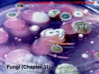
Chapter 30 Fungi and Fungi Infections H ong Xiuhua. briefs. 1. Fungi Pathogenic for Humans 2. Mycoses-the diseases Caused by Fungi 3 .Diagnostic Procedure (By Disease Type) 4. Antigenic Beochemical And Molecular Markers Antifun For Diagnosis Of Invasive Fungal Infection

E N D
briefs 1. Fungi Pathogenic for Humans 2. Mycoses-the diseases Caused by Fungi 3 .Diagnostic Procedure (By Disease Type) 4. Antigenic Beochemical And Molecular MarkersAntifun For Diagnosis Of Invasive Fungal Infection 5. Antifungal Susceptibility Testing
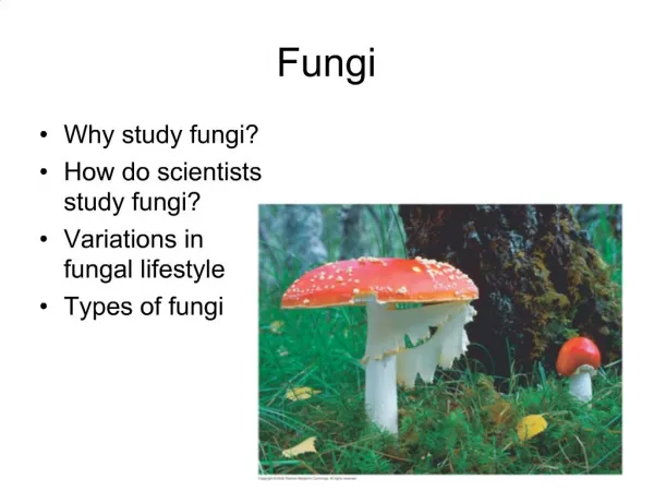
Fungi Pathogenic for Humans Fungi are eukaryotic organisms Fungi reproduction The two main growth forms of fungi
Fungi are eukaryotic organisms Fungi are classified in their own distinct taxon, the kingdom Fungi (or kingdom Mycou). Fungi are true eukaryotes having their DNA surrounded by a membrane,possess typical membrane structure and function, and have the typical array of eukaryotic organelles(e.g.,ribosomes,mitochondria). However, fungi have cell walls containing complex polysaccharides, the main ones being glucans and mannans. They also demonstrate N-acetylglucosamine or chitin in their walls.
Fungi reproduction Fungi reproduce both sexually and asexually and exist in literally thousands of different morphologic expressions and multitudinous life cycles. Most relevant to medical mycology is the ability of fungi to produce asexual propagules of many types, shapes, and size.
The two main growth forms of fungi The two main growth forms of fungi consist of single-celled, budding forms called yeasts and multicellular, filamentous forms called molds. Among medical fungi, several possess the ability to convert from one morphology to another and back again, depending on the conditions of growth and the genetics of the specific organism. Such fungi are called dimorphic because of their ability to exist in two distinct morphologic forms.
Mycoses-The diseases Caused By Fungi In discussing fungai infections, we would employ the following scheme of mycotic infection classification the scheme of mycotic infection classificationSuperficial 1.mycosesCutaneous 2.mycosesSubcutaneous 3.mycosesOpportunistic 4.mycoses Systemic 5.mycoses
Superficial mycoses These mycoses infect the superficial layer of the skin. A very common infections is pityriasis versicolor caused by the yeastlike fungus Malassezia furfur.
Cutaneous mycoses These mycoses affect the hair, skin, and nails. Dermatophytoses are caused by three genera of dermatophytes(Trychophyton, Microsporum, and Epidermatophyton).
Subcutaneous mycoses These mycoses affect not only the skin but also the subcutaneous tissues and even bone. Classic subcutaneous mycoses are sporotrichosis (Sporothrix schenckii),chromoblastomycosis(caused by at least six different species of darkly pigmented molds ) , and eumycotic mycetoma(caused by a wide variety of fungi)
Systemic(deep) mycoses The classic systemic (deep) fungal agents are dimorphic: Coccidioides immitis (coccidioidomycosis) Histoplasma capsulatum(histoplasmosis) Blastpmyces dermatitidis (blastpmycosis)
Opportunistic mycoses Opportunistic mycoses are those occurring in the host with abrogated or altered immunity. The classic Opportunistic mycoses still remain: candidiasis (largely owing to Candida albicans) cryptococcosis (Cryptococcus neoformans) aspergillosis (Aspergillus fumigatus and A.flavus) zygomycosis (Rhzopus arrhizus) phaeohyphomycosis (darkly pigmented molds and yeasts) hyalohyphomycosis (lightly pigmented,colorless or hyaline mold)
Diagnostic Procedure(By Disease Type) Superficial Mycoses Subcutaneous Mycoses Cutaneous mycoses Systemic mycoses Opportuntstic Mycoses
Superficial Mycoses KOH microscopic examination reveals a characteristic-spaghetti and meatballs appearance owing to the fragment of hyphae mixed with round yeast cells. It is the most important diagnostic procedure . At present there no direct antigen or DNA methord nor serologic procedure useful for detection of this yeast.
Cutaneous mycoses Skin scraping, nail scraping ,and observation of hairs directly by microscopy are also most helpful in diagnosing dermatophytic infetions. Employing a drop of potassium hydroxide (15%KOH ) on a glass slide over the surface of the specimen will often reveal filamentous , clear hyphal elements characteristic of dermatophytes. Calcofluor white has been employed in examining specimens for fungal elements and has demonstrated excellent results.
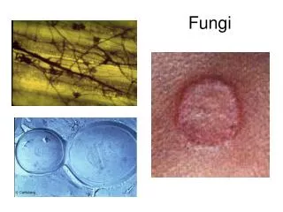
Potassium hydroxide preparation demonstrates hyphal elements characterisy ofdermatophytes.Trichophyton tonsurans in hair shaft (original magnification×600
Macrocondia and Microcondia of Microsporum cannis (original magnification×625)
produces variably sized and shaped microconidia, with relatively large spherical conidia often being right alongside of small, parallel-walled conidia and other microconidia of various sizes and shapes. Trichophyton tonsurans
Cultures(1) Cultures are always desirable and can be obtained on any number of laboratory media such as Sabouraud agar,with or without antibiotics,or dermatophyte test medium. An alternative is employment of potato flakes agar, containing the color indicator bromthymol blue .With this medium,the dermatophytic fungi change the medium from yellow to blue but do not obscure the pigments produced by various dermatophytes that are often useful as adjuncts in identification.
Cultures (2) Also,this medium promotes sporulation of dermato- phytes to enhance microscopic identification .As with dermatophyte test medium,many nondermatophytic fungi also grow on potato flakes agar and may turn the medium blue. The final identification lies with examination of the fungus under the microscope.
Subcutaneous Mycoses The most common subcutaneous mycoses: 1.lymphocutaneous sporotrichosis 2.chromoblastomycosis 3.eumycotic mycetoma
grows within 2 to 5 days on a variety of mycologic media and appears as a budding yeast at 37℃ and as a mold at 25℃. The colonies at 25℃ are initially white and moist, turning brown to black with prolonged incubation. lymphocutaneous sporotrichosis(1) S.schenickii
lymphocutaneous sporotrichosis(2) Microscopically,the mold appears as delicate branching hyphae with numerous conidia developing in a rosseIte pattern at the ends of conidiophores. Laboratory confirmation is establishied by converting the mycelial growth to the yeast form by subculture at 37℃. Alternatively,the organism may be identified immunologically by using the exoantigen test
chromoblastomycosis The diagnosis of chromoblastomycosis is generally made by histopathologicexamination of infected tissue. Typical lesions demonstrate pseudoepitheliomatous hyperplasia and characteristic brown or copper-colored spherical cells or hyphae known as sclerotic or Medlar bodies. The cultures grow as dematiaceous mold but may take weeks to appear and longer to develop the characteristic conidia.
eumycotic mycetoma(1) Examination of the draing from sinus(eumycotic mycetoma) tracts may reveal small granules or microcolonies of mycelial filaments.These granules may range from microscopic to 2 mm in diameter and vary according to the infecting species Histopathologic examination of tissue may reveal hyphal elements and granules. Identification of the fungi causing eumycotic mycetomas is by morphology of the asexual conidia formed in culture.
eumycotic mycetoma(2) filaments.These granules may range from microscopic to 2 mm in diameter and vary according to the infecting species .Histopathologic examination of tissue may reveal hyphal elements and granules. Identification of the fungi causing eumycotic mycetomas is by morphology of the asexual conidia formed in culture
Systemic mycoses owing to Dimorphic Fungal Pathogen The dimorphic: fungal pathogen are organism that exist in a mold form in natural environment in the laboratory at 250c to 300c and in the yeast or spherule form in tissues or when grown on enriched medium in the laboratory at 370c.
Systemic mycoses owing to Dimorphic Fungal Pathogen Histoplasmosis Blastomycosis Coccidioidomycosis Paracoccidjoidomycosis Penicillosis
Histoplasmosis(1) Histoplasma capsulatum var capsulatum is most often recoverd from respiratory secretions or from blood or bone marrow. It is recommended that the specimen be placed directly on BHI medium with chloramphenicol and gentamicin . When an organism is isolated,it should be transferred to potato flakes, Sabhi, or Gormans media.
When H. capsulatum is suspected further laboratory testing is required to diffrentiate it from saprobic fungi. Conversion to the yeast form is achieved by incubating the culture at 370C on BHI agar. Histoplasmosis(2)
Histoplasmosis(3) Other more rapid means of identifying H. capsulatum from culture include the exoantigen test and the use of DNA probes. Additional means of diagnosing histoplasmosis includes histopathologic examination of infected tissue and serology. Serologic diagnosis employs both ID and complement fixation (CF)tests and has been quite useful.
Blastomycosis(1) When blastomycosisis suspected,clinical material (e.g.,sputum,pus)should be placed on inhibitory media, without cycloheximide, such as BHI agar with chloramphenicol and gentamicin and incubated at 25℃ for 7 to 28 days. Microscopic examination at this points not diagnostic. .
Blastomycosis(2) Probable Blastomyces isolates must be converted to the yeast phase by subculturing the isolate to a BHI blood agar slant and incubating at 37℃ in.Yeast forms should be visible within a week. Microscopic examination at this point reveals characteristic broad-based budding yeasts.
Although microscopic examination of the yeast gives valuable information,a definitive identification may be made from mycelial phase cultures using the exoantigen test as described for H.capsulatum. A commercially available DNA probe assay also allows the rapid identification (2h)of this organism from culture.Additional means of diagnosing blastomycosis includes histopathologic examination of infected tissue. Blastomycosis(3)
Blastomycosis(4) Serologic tests for diagnosing blastomycosis have not been particularly useful. Antigens for CF tests are available,but complement fixing antibodies are absent in as many as 50%of cases.
The tissue phase of B.dermdtitidisis a large yeast with broad-based buds
Coccidioidomycosis(1) C.immitis is a dimorphic fungus with a variety of mold morphologies at 25℃. Initial growth is white to gray,moist,and glabrous and occurs within 3 to 4 days. It rapidly develops abundant aerial mycelia,and the colony appears to enlarge in a circular "bloom”. Mature colonies usually become tan to brown to lavender.
Identification of C. imminitis from culture may be accomplished by using the exoantigen or DNA probe tests. Additional means of diagnosing coccidioidomycosis include histopathologic examination of infected tissue for the presence of spherules and serologic testing. Coccidioidomycosis(2)
Coccidioidomycosis(3) Several serologic procedures exist for initial screening,confirmation,and prognostic evaluation.For initial diagnosis,the combined use of the immmunodiffusion(ID) test and the latex particle agglutination test detects approximately 93% of cases. The CF and tube precipitin tests may also be employed for diagnosis as well as for confirmation.
Paracoccidjoidomycosis(1) P.brasiliensis is a dimorphic fungus that produces a variety of mold morphologies when grown at 25℃.Flat colonies are glabrous to leathery, wrinkled to folded, floccose to velvety, pink to beige to brown,with a yellowish-brown reverse, and resemble those of B.dermdtitis . Diagnosis may be made by culture, histopathologic examination of infected tissues,and serology.BothOn BHI blood agar at 37 ℃,the mycelial phase rapidly converts to the yeast phase.
CF and agar gel ID procedures are available for serodiagnosis.The CF test,using yeast-derived antigen,is positive in 80% of active disease.Titers of l:64 or higher are generally considered diagnostic .Although the CF titer usually decreases with therapy,it may persist in the range of 1:8 to 1:32.The ID test is positive in approximately 95% of active cases,has low cross,reactivity,and diminishes with successful therapy. Paracoccidjoidomycosis(3)
Penicillosis(1) Most infection of the Penicillum marneffei occur in HIV-infected hosts. Detection of Penicillum marneffei by histopathologic examination or by culture of blood,bone marrow,skin,and respiratory specimens is the most productive means of making a laboratory diagnosis of infection with this organism .In tissue,the presence of 4,t0 8μm diameter cells that divide by fission is characteristic of P. marneffeii.
Penicillosis(2) In culture, P. marneffeii produces a diffuse red pigment and grows as a mold at 25 ℃ to 30 ℃. When incubated at 37 ℃, it undergoes a phase transition and grows as a single celled yeast form. The red pigment is not produced by the yeast phase o f the organism.
Opportuntstic Mycoses The most well-known causes of opportunistic mycosesCandida and Other Opportunistic Yeasts Aspergillosis Zygomycosis