Smooth Muscle
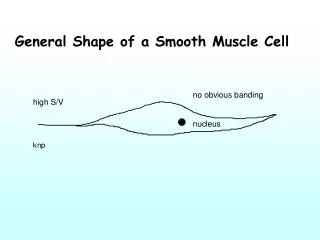
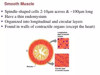
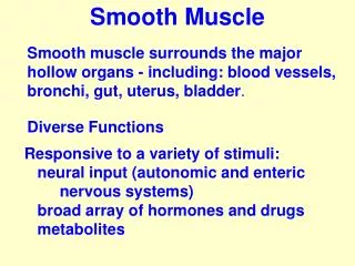
Smooth Muscle. Characteristics Not striated Dense bodies instead of Z disks as in skeletal muscle Have noncontractile intermediate filaments Ca 2+ required to initiate contractions Types Visceral or unitary Function as a unit Multiunit Cells or groups of cells act as independent units.

Smooth Muscle
E N D
Presentation Transcript
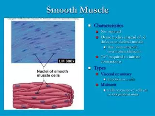
Smooth Muscle • Characteristics • Not striated • Dense bodies instead of Z disks as in skeletal muscle • Have noncontractile intermediate filaments • Ca2+ required to initiate contractions • Types • Visceral or unitary • Function as a unit • Multiunit • Cells or groups of cells act as independent units
Functional Properties of Smooth Muscle • Some visceral muscle exhibits autorhythmic contractions • Tends to contract in response to sudden stretch but no to slow increase in length • Exhibits relatively constant tension: Smooth muscle tone • Amplitude of contraction remains constant although muscle length varies
Smooth Muscle Regulation • Innervated by autonomic nervous system • Neurotransmitter are acetylcholine and norepinephrine • Hormones important as epinephrine and oxytocin • Receptors present on plasma membrane which neurotransmitters or hormones bind determines response
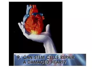
Cardiac Muscle • Found only in heart • Striated • Each cell usually has one nucleus • Has intercalated disks and gap junctions • Autorhythmic cells • Action potentials of longer duration and longer refractory period • Ca2+ regulates contraction
Cardiac Muscle • Elongated, branching cells containing 1-2 centrally located nuclei • Contains actin and myosin myofilaments • Intercalated disks: Specialized cell-cell contacts • Desmosomes hold cells together and gap junctions allow action potentials • Electrically, cardiac muscle behaves as single unit
Refractory Period • Absolute: Cardiac muscle cell completely insensitive to further stimulation • Relative: Cell exhibits reduced sensitivity to additional stimulation • Long refractory period prevents tetanic contractions
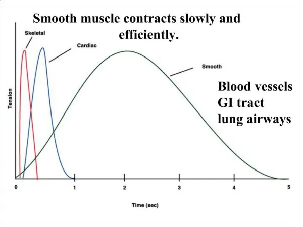
AP-contraction relationship: • AP in skeletal muscle is very short-lived • AP is basically over before an increase in muscle tension can be measured. • AP in cardiac muscle is very long-lived • AP has an extra component, which extends the duration. • The contraction is almost over before the action potential has finished.
Functions of the Heart • Generating blood pressure • Routing blood • Heart separates pulmonary and systemic circulations • Ensuring one-way blood flow • Heart valves ensure one-way flow • Regulating blood supply • Changes in contraction rate and force match blood delivery to changing metabolic needs
Orientation of cardiac muscle fibres: • Unlike skeletal muscles, cardiac muscles have to contract in more than one direction. • Cardiac muscle cells are striated, meaning they will only contract along their long axis. • In order to get contraction in two axis, the fibres wrap around.
Heart Wall • Three layers of tissue • Epicardium: This serous membrane of smooth outer surface of heart • Myocardium: Middle layer composed of cardiac muscle cell and responsibility for heart contracting • Endocardium: Smooth inner surface of heart chambers
Heart Sounds • First heart sound or “lubb” • Atrioventricular valves and surrounding fluid vibrations as valves close at beginning of ventricular systole • Second heart sound or “dupp” • Results from closure of aortic and pulmonary semilunar valves at beginning of ventricular diastole, lasts longer • Third heart sound (occasional) • Caused by turbulent blood flow into ventricles and detected near end of first one-third of diastole
Cardiac Arrhythmias • Tachycardia: Heart rate in excess of 100bpm • Bradycardia: Heart rate less than 60 bpm • Sinus arrhythmia: Heart rate varies 5% during respiratory cycle and up to 30% during deep respiration • Premature atrial contractions: Occasional shortened intervals between one contraction and succeeding, frequently occurs in healthy people
Mean Arterial Pressure (MAP) • Average blood pressure in aorta • MAP=CO x PR • CO is amount of blood pumped by heart per minute • CO=SV x HR • SV: Stroke volume of blood pumped during each heart beat • HR: Heart rate or number of times heart beats per minute • Cardiac reserve: Difference between CO at rest and maximum CO • PR is total resistance against which blood must be pumped
Cardiac Cycle • Heart is two pumps that work together, right and left half • Repetitive contraction (systole) and relaxation (diastole) of heart chambers • Blood moves through circulatory system from areas of higher to lower pressure. • Contraction of heart produces the pressure
Regulation of the Heart • Intrinsic regulation: Results from normal functional characteristics, not on neural or hormonal regulation • Starling’s law of the heart • Extrinsic regulation: Involves neural and hormonal control • Parasympathetic stimulation • Supplied by vagus nerve, decreases heart rate, acetylcholine secreted • Sympathetic stimulation • Supplied by cardiac nerves, increases heart rate and force of contraction, epinephrine and norepinephrine released
Heart Homeostasis • Effect of blood pressure • Baroreceptors monitor blood pressure • Effect of pH, carbon dioxide, oxygen • Chemoreceptors monitor • Effect of extracellular ion concentration • Increase or decrease in extracellular K+ decreases heart rate • Effect of body temperature • Heart rate increases when body temperature increases, heart rate decreases when body temperature decreases